
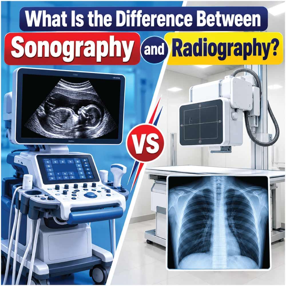
The difference between sonography and radiography mainly comes down to how medical images are produced, what conditions they are commonly used to examine, and what the imaging professional does during the procedure. Both are important diagnostic imaging fields, but they rely on different technologies and require different technical skills.
Sonography uses high-frequency sound waves to create images of organs, tissues, blood vessels, and developing fetuses. Radiography, by contrast, uses X-rays to produce images, particularly of bones, the chest, and other structures that can be visualized effectively with X-ray equipment.
The two professions also differ in their daily workflow. A sonographer generally works closely with the patient while manipulating a handheld transducer and obtaining images in real time. A radiographer positions the patient and operates X-ray equipment to produce diagnostic images while applying radiation-safety principles.
For someone considering a healthcare career, understanding these distinctions is useful because the two paths can lead to different clinical environments, specialties, equipment, and professional responsibilities.
Sonography Explained
Sonography, also called diagnostic medical ultrasound, creates images by sending high-frequency sound waves into the body and analyzing the returning echoes. The resulting information is displayed as images on an ultrasound monitor.
A sonographer typically applies ultrasound gel to the patient’s skin and moves a transducer over the examination area. The transducer sends sound waves into the body and receives the returning signals. The sonographer then adjusts the equipment and captures images that are useful for the interpreting physician.
Unlike X-ray imaging, diagnostic sonography uses nonionizing sound waves rather than ionizing radiation.
Sonography can be used for many types of examinations, including:
- Abdominal ultrasound
- Obstetric and fetal imaging
- Pelvic ultrasound
- Vascular studies
- Breast ultrasound
- Cardiac imaging
- Musculoskeletal ultrasound
- Other specialized examinations
Sonographers may work in hospitals, outpatient imaging centers, physician offices, and diagnostic clinics.
Radiography Explained
Radiography is the medical imaging specialty most closely associated with conventional X-rays. A radiographer operates X-ray equipment to capture images of structures such as bones, the chest, and internal organs.
The professional must position the patient correctly, select appropriate technical settings, operate the equipment, and produce images that meet diagnostic requirements. Radiographers may also assist with certain procedures, including fluoroscopic examinations and studies involving contrast media.
Radiography is especially valuable when clinicians need fast images of:
- Fractures and other bone injuries
- The chest and lungs
- Certain abdominal conditions
- Joint abnormalities
- Foreign objects
- Other conditions that can be assessed effectively with X-ray imaging
Radiographers can also work with other imaging technologies as they gain additional education and credentials.
Sonography vs Radiography: Key Differences
Although both professions are part of diagnostic medical imaging, their technologies and clinical workflows are substantially different.
| Feature | Sonography | Radiography |
|---|---|---|
| Primary technology | High-frequency sound waves | X-rays |
| Ionizing radiation | No | Yes |
| Main equipment | Ultrasound system and transducer | X-ray system |
| Image production | Echoes from sound waves | X-ray absorption patterns |
| Patient interaction | Often continuous during scanning | Usually focused on positioning and imaging |
| Common applications | Soft tissues, vessels, pregnancy, organs | Bones, chest, and many general radiographic exams |
| Real-time imaging | Yes | Generally image acquisition rather than continuous ultrasound-style scanning |
| Common work environments | Hospitals, clinics, imaging centers, physician offices | Hospitals, imaging centers, clinics, emergency departments |
The distinction is not simply that one profession uses “ultrasound” and the other uses “X-rays.” The physical principles, equipment operation, image acquisition process, and clinical responsibilities are different.
How the Imaging Technology Differs
How Sonography Produces an Image
Ultrasound imaging depends on sound waves. The transducer sends pulses into the body and receives echoes as those waves encounter tissues with different acoustic properties.
The ultrasound system processes these returning signals to construct an image. Because the transducer can be moved across the body, the sonographer can examine anatomy from different angles and observe structures dynamically.
This real-time capability is one of the defining characteristics of sonography.
For example, during an abdominal examination, a sonographer may adjust the transducer position, pressure, angle, and imaging settings to obtain useful views of different organs.
How Radiography Produces an Image
Radiography uses X-rays, which are a form of ionizing radiation. The X-ray beam passes through the body, and different tissues absorb different amounts of radiation.
The resulting pattern is recorded by a detector and processed into an image.
Bone absorbs substantially more X-rays than many surrounding soft tissues, which is why skeletal structures are prominently displayed on conventional radiographs.
Because radiography involves ionizing radiation, radiation protection is an important part of professional practice. Radiologic technologists use safety techniques designed to keep unnecessary exposure as low as reasonably achievable.
Which One Uses Radiation?
One of the most important differences between sonography and radiography is radiation exposure.
Diagnostic sonography does not use ionizing radiation. It uses high-frequency sound waves to create images.
Radiography uses X-rays, which are ionizing radiation. Consequently, radiographers must understand radiation safety and apply appropriate protective practices during examinations.
This difference can influence which imaging method a healthcare provider selects for a particular clinical question.
However, it does not mean that one imaging method can replace the other. Ultrasound and X-ray imaging provide different types of information and are selected according to the patient’s condition, the anatomy being examined, and the diagnostic question.
What Does a Sonographer Do?
A sonographer has responsibilities that go beyond simply operating an ultrasound machine.
During an examination, the professional may:
- Review the examination request and prepare the patient.
- Explain the procedure.
- Position the patient appropriately.
- Apply ultrasound gel.
- Operate and adjust ultrasound equipment.
- Move the transducer across the examination area.
- Capture diagnostic images and measurements.
- Optimize image quality.
- Document relevant findings according to professional procedures.
- Communicate appropriately with the interpreting physician and healthcare team.
Sonographers are therefore highly involved in the image-acquisition process. Their technical ability to recognize anatomy and obtain useful views is an important part of producing a diagnostically valuable examination.
Depending on their training, sonographers may specialize in areas such as abdominal, obstetric, vascular, cardiac, breast, or other forms of ultrasound imaging.
What Does a Radiographer Do?
Radiographers are responsible for producing diagnostic radiographic images using X-ray equipment.
Typical responsibilities include:
- Preparing and positioning patients.
- Selecting appropriate imaging techniques.
- Operating radiographic equipment.
- Adjusting exposure-related technical factors.
- Capturing diagnostic images.
- Checking images for technical quality.
- Applying radiation-safety practices.
- Using protective equipment and appropriate positioning when required.
- Communicating with patients and members of the healthcare team.
Radiographers may also work with mobile X-ray equipment in locations such as emergency departments, operating rooms, or patient rooms.
Their role requires a combination of anatomy knowledge, technical equipment skills, patient-care abilities, and radiation-safety knowledge.
Education and Training Differences
The education required depends on the country, credentialing organization, employer, and specialty. In the United States, both radiography and sonography can be entered through formal educational programs followed by professional credentialing.
ARRT lists both Radiography and Sonography among disciplines available through its primary eligibility pathway. Its primary pathway requires an associate degree or higher and completion of an ARRT-approved educational program in the discipline being pursued.
Radiography programs generally include subjects such as:
- Human anatomy
- Patient care
- Radiographic procedures
- Radiation physics
- Radiation protection
- Image production
- Equipment operation
- Clinical practice
Sonography education focuses on areas such as:
- Anatomy and physiology
- Ultrasound physics
- Patient care
- Image optimization
- Scanning techniques
- Pathology
- Clinical ultrasound experience
- Specialty-specific examinations
ARRT’s requirements include both didactic education and clinical competency requirements for its credentials.
For sonography, ARDMS also has specific education and clinical-experience pathways for candidates seeking its credentials. Requirements vary according to the prerequisite pathway used.
Certification and Professional Credentials
Certification is another area where the two career paths can differ.
In the United States, ARRT offers credentials in both radiography and sonography. Candidates must meet the applicable education, ethics, and examination requirements.
For ultrasound professionals, ARDMS is another major credentialing organization. Its credentials cover areas of diagnostic medical sonography and related specialties.
The exact credential required or preferred by an employer can vary, so students should check the requirements of the hospitals, clinics, and states where they intend to work.
Professional requirements can also change over time. Anyone choosing a program should verify current eligibility requirements directly with the relevant credentialing organization before enrolling.
Also Read:
Where Do Sonographers and Radiographers Work?
Both professionals can work in hospitals and outpatient healthcare settings, but their daily environments may differ depending on their specialty.
| Work setting | Sonography | Radiography |
|---|---|---|
| Hospitals | ✓ | ✓ |
| Outpatient imaging centers | ✓ | ✓ |
| Physician offices | ✓ | ✓ |
| Emergency departments | Sometimes | ✓ |
| Bedside/mobile imaging | ✓ | ✓ |
| Specialized cardiac services | ✓ | — |
| General X-ray department | — | ✓ |
| Vascular imaging | ✓ | — |
The actual workplace mix depends on local healthcare systems, employer needs, credentialing, and the professional’s specialty.
Sonography Specialties vs Radiography Specialties
Sonography can branch into several specialized areas.
Common Sonography Specialties
Examples include:
- Abdominal sonography: Imaging abdominal organs and related structures.
- Obstetric sonography: Imaging pregnancy and fetal development.
- Vascular sonography: Evaluating blood vessels and blood flow.
- Cardiac sonography: Producing ultrasound images of the heart.
- Breast sonography: Examining breast tissue using ultrasound.
- Musculoskeletal sonography: Evaluating selected muscles, tendons, ligaments, and other structures.
Radiography can also provide a foundation for further imaging specialization.
Common Radiography-Related Paths
Radiography professionals may later pursue areas such as:
- Computed tomography (CT)
- Magnetic resonance imaging (MRI)
- Mammography
- Interventional radiography
- Bone densitometry
- Other advanced imaging areas
ARRT’s postprimary pathways demonstrate how radiography can serve as a supporting credential for several advanced disciplines, depending on the credential and requirements.
Also Read:
How Patient Interaction Differs
Patient communication is important in both careers, but the nature of the interaction can be different.
A sonographer may spend a considerable amount of time directly interacting with a patient during an examination. The professional may continuously reposition the transducer, ask the patient to change position or hold their breath, and make adjustments based on the anatomy being examined.
Radiographers also interact directly with patients, but many examinations can be completed relatively quickly once the patient is correctly positioned.
Both roles require:
- Clear communication
- Respect for patient privacy
- Safe positioning
- Professional behavior
- Attention to patient comfort
- Awareness of mobility limitations and clinical conditions
Strong patient-care skills can therefore be valuable in either profession.
Also Read:
Which Career Requires More Technical Skill?
Both careers require substantial technical knowledge, but the nature of that technical skill is different.
Sonography can require extensive hand-eye coordination because the professional must manipulate the transducer while watching the image and recognizing anatomy in real time. The sonographer also has to select appropriate views and optimize the examination.
Radiography requires precise positioning, equipment operation, technical-factor selection, image evaluation, and radiation-safety practices.
Rather than viewing one profession as technically harder than the other, it is more useful to consider the type of work involved.
| Skill area | Sonography | Radiography |
|---|---|---|
| Anatomy knowledge | High | High |
| Patient positioning | High | High |
| Equipment operation | High | High |
| Real-time scanning | High | Low |
| Radiation safety | Not based on ionizing radiation | Essential |
| Hand-eye coordination | High | Important |
| Image-quality evaluation | High | High |
| Patient communication | High | High |
Sonography vs Radiography: Choosing a Career Path
Someone considering these careers should look beyond the equipment and think about what the day-to-day work actually involves.
Sonography may appeal to people who enjoy:
- Real-time imaging
- Close patient interaction
- Anatomy-based problem solving
- Hands-on scanning
- Specialized ultrasound examinations
- Working without ionizing radiation
Radiography may appeal to people interested in:
- X-ray imaging
- Bone and chest examinations
- Fast-paced imaging environments
- Equipment and positioning
- Emergency and hospital imaging
- Opportunities to build toward other imaging specialties
Neither description applies identically to every workplace. Clinical environments, schedules, patient populations, and responsibilities vary considerably.
Can a Radiographer Become a Sonographer?
In some credentialing systems, professionals can build additional qualifications after completing an initial imaging credential.
ARRT has pathways that recognize existing credentials when candidates pursue certain additional disciplines. For example, ARRT states that an ARRT or ARDMS sonography credential can serve as a supporting credential for selected postprimary pathways, while radiography can serve as a supporting category for several other imaging credentials.
However, becoming qualified in sonography is not simply a matter of learning how to operate an ultrasound machine. Formal education, clinical competency, examinations, and credential-specific requirements may apply.
Anyone considering a transition should check the exact requirements for the credential and jurisdiction they intend to use.
Similarities Between Sonography and Radiography
Despite their differences, the professions have a lot in common.
Both professionals:
- Work in healthcare environments.
- Produce diagnostic images for physicians and other providers.
- Need strong anatomy knowledge.
- Work directly with patients.
- Operate specialized medical equipment.
- Must maintain image quality.
- Follow patient-safety procedures.
- Require formal education and clinical training.
- Can pursue additional specialties.
Both careers also contribute to a larger diagnostic process. The imaging professional obtains technically appropriate images, while the interpreting physician evaluates the images and incorporates them into the patient’s clinical assessment.
Sonography vs Radiography: Quick Comparison
| Category | Sonography | Radiography |
|---|---|---|
| Imaging energy | Sound waves | X-rays |
| Ionizing radiation | No | Yes |
| Main professional | Sonographer | Radiographer/radiologic technologist |
| Main equipment | Ultrasound machine and transducer | X-ray system and detector |
| Real-time scanning | Yes | Generally no |
| Strong application areas | Soft tissue, vessels, pregnancy, organs | Bones, chest, general radiographic imaging |
| Patient contact | Often extensive during scan | Significant during positioning and examination |
| Radiation protection | Not applicable to ionizing radiation | Major responsibility |
| Career specialization | Abdominal, vascular, cardiac, obstetric, breast, etc. | CT, MRI, mammography, interventional, etc. |
| Credentialing | ARDMS, ARRT, depending on pathway | ARRT and applicable state/employer requirements |
Final Thoughts on the Difference Between Sonography and Radiography
The difference between sonography and radiography is primarily rooted in imaging technology and professional workflow. Sonography uses high-frequency sound waves and produces images in real time, while radiography uses X-rays to create diagnostic radiographs.
The distinction also extends to education, equipment, patient interaction, safety practices, and specialty opportunities.
Sonographers spend much of their time acquiring and optimizing ultrasound images through hands-on scanning. Radiographers focus on positioning patients and operating X-ray equipment while applying strict radiation-safety principles.
Both professions require technical knowledge, clinical training, patient-care skills, and attention to image quality. The right path depends on the type of imaging technology, clinical environment, patient interaction, and professional specialization that best fits an individual’s interests and career plans.
Because certification and licensing requirements can vary by location, prospective students should always confirm current requirements with the appropriate credentialing organization and local authorities before selecting an educational program.

Zak is a dedicated medical and career writer specializing in sonography, healthcare education, and professional development. Through SonographerSalary.com, he shares in-depth insights on sonographer salaries, education pathways, and career tips to help readers build successful futures in medical imaging. His content combines accuracy with practical, easy-to-understand guidance, empowering students and professionals to make confident, informed career decisions.