
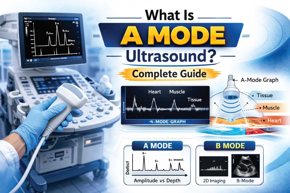
A Mode ultrasound, or Amplitude Mode ultrasound, is one of the earliest types of diagnostic sonography. It represents tissue structures as spikes along a single line, showing echo amplitude and depth. This mode provides one-dimensional imaging, forming the basis for more advanced ultrasound techniques.
A-Mode ultrasound is primarily used in specialized fields such as ophthalmology and some vascular assessments. Although less common today, it remains essential for precise measurements of distances and tissue interfaces. Its simplicity and high accuracy make it useful in select clinical applications.
Understanding the Basics of A-Mode Ultrasound
A-Mode stands for Amplitude Mode, where the echo strength is displayed as a spike along the vertical axis. The horizontal axis represents the depth of tissue, measured from the transducer. The height of each spike corresponds to the strength of the reflected sound wave.
This mode does not produce a two-dimensional image like B-Mode. Instead, it focuses on tissue reflectivity and distance measurements, providing highly precise structural information in a single dimension. It is particularly useful in applications requiring exact depth calculations.
How A-Mode Ultrasound Works
A-Mode ultrasound operates by sending high-frequency sound waves into the body through a transducer. Echoes from tissue interfaces return to the transducer, generating spikes on a graph. The spike height indicates echo amplitude, and the spike location indicates tissue depth.
The system records these reflections along a single scan line. By analyzing the amplitude and position of each spike, clinicians can determine tissue boundaries, measure distances between structures, and evaluate specific anatomical features with high accuracy.
Key Components of an A-Mode Ultrasound System
| Component | Function |
|---|---|
| Transducer | Emits and receives ultrasound waves |
| Pulser | Generates the electrical pulses for sound waves |
| Receiver | Detects returning echoes |
| Display Monitor | Shows the amplitude spikes along depth |
| Measurement Software | Calculates distances between echo spikes |
Each component ensures precise signal acquisition, accurate display, and effective measurement of tissue interfaces in one-dimensional ultrasound imaging.
Image Formation in A-Mode Ultrasound
The A-Mode display is a spike graph, not a two-dimensional image. The vertical axis shows echo amplitude, while the horizontal axis represents distance from the transducer. Strong reflections create taller spikes, weak reflections produce smaller ones.
For example, in ocular imaging, the cornea, lens, and retina create distinct spikes. By measuring spike positions, ophthalmologists can calculate axial eye length, detect tumors, and assess other structural parameters with remarkable precision.
Frequency Ranges Used in A-Mode Ultrasound
| Frequency Range | Penetration | Resolution | Common Uses |
|---|---|---|---|
| 5 – 10 MHz | Medium | High | Eye measurements, small organs |
| 10 – 20 MHz | Low | Very high | Ophthalmology, corneal imaging |
| 2 – 5 MHz | High | Moderate | Certain vascular measurements |
Higher frequencies offer better resolution but reduced penetration, making frequency selection crucial for accurate imaging of specific structures.
Clinical Applications of A-Mode Ultrasound
1- Ophthalmology
A-Mode is widely used to measure axial length of the eye, corneal thickness, and lens position. These measurements are critical for cataract surgery planning, intraocular lens placement, and detection of ocular abnormalities.
2- Vascular Assessment
A-Mode can assist in evaluating vessel wall thickness and certain vascular pathologies. Though less common than B-Mode, it provides precise measurements where distance accuracy is critical.
3– Oncology
In specific cases, A-Mode helps detect tumor margins by analyzing echo spikes from tissue interfaces, providing quantitative data for treatment planning and surgical guidance.
Read More:
Advantages of A-Mode Ultrasound
| Advantage | Explanation |
|---|---|
| High measurement accuracy | Ideal for precise distance calculations |
| Simple operation | Easy to use with basic equipment |
| Rapid acquisition | Quick one-dimensional imaging |
| Safe | Non-ionizing sound waves, suitable for repeated use |
| Useful in specialized applications | Ophthalmology, certain vascular assessments |
Despite its simplicity, A-Mode remains valuable in clinical scenarios where distance accuracy is more critical than anatomical visualization.
Limitations of A-Mode Ultrasound
| Limitation | Reason |
|---|---|
| One-dimensional image | Cannot display anatomical context |
| Limited applications | Primarily ophthalmology and select vascular use |
| Operator-dependent | Requires precise transducer alignment |
| Less informative than B-Mode | Cannot visualize organ structures clearly |
A-Mode is rarely used alone today but still plays a supportive role in combination with B-Mode or other modalities.
A-Mode vs B-Mode Ultrasound
| Feature | A-Mode | B-Mode |
|---|---|---|
| Image type | One-dimensional spikes | 2D grayscale image |
| Axis | Vertical → amplitude, horizontal → depth | Both axes → spatial representation |
| Main use | Distance measurements | Anatomical imaging |
| Temporal resolution | High | Moderate |
| Common application | Ophthalmology | General imaging (abdominal, cardiac, vascular) |
A-Mode provides quantitative precision, while B-Mode offers visual context; together, they complement modern ultrasound examinations.
Read More:
Role of A-Mode in Modern Sonography
Although modern sonography largely relies on B-Mode, M-Mode, and Doppler imaging, A-Mode still has relevance in specialized quantitative applications. It is particularly critical when precise linear measurements are needed.
In ophthalmology, A-Mode is still the standard for calculating intraocular lens power and measuring axial eye length, forming the foundation for refractive surgery planning.
Preparation for an A-Mode Ultrasound Scan
Preparation varies by application. For ocular imaging, patients simply rest their head while the probe is gently placed against the eye using a coupling gel. No fasting or invasive preparation is needed.
For vascular measurements, minimal preparation is required. Proper probe placement and patient cooperation ensure accurate distance readings. Operator expertise is essential for obtaining reliable and reproducible measurements.
Safety of A-Mode Ultrasound
A-Mode is extremely safe because it uses non-ionizing ultrasound waves. It is suitable for repeated use, including in sensitive structures like the eye. No known side effects have been reported when performed within standard clinical guidelines.
Its safety profile, combined with high measurement accuracy, ensures continued relevance in ophthalmology and specialized diagnostic procedures.
Learning A-Mode for Sonography Students
For students of diagnostic sonography, understanding A-Mode is essential because it:
- Forms the basis for B-Mode and M-Mode
- Teaches principles of ultrasound wave reflection and echo amplitude
- Demonstrates how measurement accuracy is achieved using ultrasound
Students must learn proper probe alignment, signal interpretation, and distance measurement techniques for accurate results.
Read More:
Future of A-Mode Ultrasound
Though largely replaced by B-Mode and M-Mode for general imaging, A-Mode continues evolving:
- Integration with digital measurement systems
- AI-assisted spike interpretation
- Combination with B-Mode for hybrid imaging
These advancements increase measurement reliability and reduce operator dependency, preserving A-Mode’s role in precision-focused clinical applications.
Conclusion
A-Mode ultrasound is the original ultrasound mode that uses amplitude spikes to display one-dimensional tissue information. While its applications are limited compared to modern B-Mode or M-Mode, it remains indispensable in ophthalmology, precise distance measurements, and certain vascular assessments.
Its simplicity, safety, and accuracy make it a valuable tool for specific clinical needs. Understanding A-Mode provides a foundation for learning other ultrasound modes, ensuring precise measurement techniques and deeper insight into tissue interfaces.
A-Mode may not be widely used in general imaging today, but in specialized applications, it continues to play a critical and irreplaceable role in diagnostic sonography.

Zak is a dedicated medical and career writer specializing in sonography, healthcare education, and professional development. Through SonographerSalary.com, he shares in-depth insights on sonographer salaries, education pathways, and career tips to help readers build successful futures in medical imaging. His content combines accuracy with practical, easy-to-understand guidance, empowering students and professionals to make confident, informed career decisions.