
B Mode ultrasound, also known as Brightness Mode ultrasound, is one of the most commonly used imaging techniques in modern diagnostic medical sonography. It provides a real-time, two-dimensional grayscale image of internal body structures and plays a central role in detecting, evaluating, and monitoring a wide range of medical conditions.
From abdominal scans to obstetrics, cardiology, and vascular imaging, B-Mode ultrasound is considered the foundation of ultrasound technology. In this complete guide, you will learn how it works, its applications, advantages, limitations, and how it compares with other ultrasound modes.
Understanding the Basic Concept of B-Mode Ultrasound
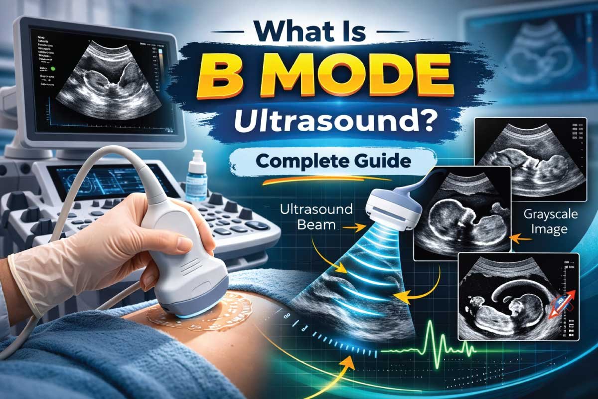
B-Mode ultrasound is based on the transmission of high-frequency sound waves into the body using a transducer. These sound waves reflect back when they encounter tissues, organs, or fluids.
The machine then converts these returning echoes into a grayscale image, where:
- Strong echoes appear bright (white)
- Weak echoes appear dark (black)
- Fluid-filled structures appear anechoic (completely black)
This brightness variation is the reason it is called Brightness Mode (B-Mode).
Unlike A-Mode, which shows amplitude spikes, B-Mode produces a detailed 2D cross-sectional image, making it ideal for anatomical visualization.
How B-Mode Ultrasound Works
The working mechanism of B-Mode ultrasound involves several steps:
- The transducer emits sound waves into the body
- Sound waves interact with tissues
- Echoes return to the transducer
- The system processes echo strength
- A real-time grayscale image is formed
This process happens within milliseconds, allowing live imaging of moving structures such as the beating heart or fetal movements.
Key Components of a B-Mode Ultrasound System
| Component | Function |
|---|---|
| Transducer | Sends and receives sound waves |
| Pulser | Generates electrical pulses |
| Receiver | Detects returning echoes |
| Scan Converter | Converts signals into image format |
| Display Monitor | Shows the grayscale image |
Image Formation in B-Mode Ultrasound
The image in B-Mode is formed using:
- Echo amplitude → brightness
- Time of return → depth
- Scanning lines → 2D image
This allows clinicians to evaluate:
- Organ size
- Tissue texture
- Lesions or masses
- Fluid accumulation
Frequency Ranges Used in B-Mode Ultrasound
Different clinical applications require different ultrasound frequencies.
| Frequency Range | Penetration | Resolution | Common Uses |
|---|---|---|---|
| 2 – 5 MHz | High | Moderate | Abdomen, obstetrics |
| 5 – 10 MHz | Medium | High | Pelvic organs, vascular |
| 10 – 18 MHz | Low | Very high | Thyroid, breast, MSK, superficial structures |
Lower frequencies penetrate deeper, while higher frequencies provide better resolution.
Clinical Applications of B-Mode Ultrasound
B-Mode ultrasound is widely used in almost every medical imaging department.
1. Abdominal Imaging
It helps evaluate:
- Liver
- Gallbladder
- Kidneys
- Pancreas
- Spleen
It is commonly used to detect:
- Gallstones
- Tumors
- Cysts
- Fatty liver
2. Obstetrics and Gynecology
In obstetrics, B-Mode ultrasound is essential for:
- Fetal growth monitoring
- Placental localization
- Amniotic fluid assessment
- Detection of congenital anomalies
In gynecology, it evaluates:
- Uterus
- Endometrium
- Ovaries
3. Cardiac Imaging (Echocardiography)
B-Mode provides structural information about:
- Heart chambers
- Valve morphology
- Wall motion
It is usually combined with M-Mode and Doppler for complete cardiac assessment.
4. Vascular Imaging
It helps visualize:
- Vessel walls
- Plaque formation
- Thrombus
5. Musculoskeletal Imaging
Used to assess:
- Tendons
- Ligaments
- Muscles
- Joint effusion
Advantages of B-Mode Ultrasound
| Advantage | Explanation |
|---|---|
| Non-invasive | No surgery or needles required |
| Radiation-free | Safe for fetus and repeated use |
| Real-time imaging | Shows motion instantly |
| Portable | Can be used at bedside |
| Cost-effective | Cheaper than CT and MRI |
Limitations of B-Mode Ultrasound
Despite its wide use, B-Mode ultrasound has some limitations:
| Limitation | Reason |
|---|---|
| Operator dependent | Image quality depends on sonographer skill |
| Limited penetration in obese patients | Sound attenuation |
| Poor imaging through bone and air | Strong reflection of sound waves |
| Small field of view | Compared to CT/MRI |
B-Mode vs Other Ultrasound Modes
| Mode | Full Name | Display Type | Main Use |
|---|---|---|---|
| A-Mode | Amplitude Mode | Spikes | Ophthalmology |
| B-Mode | Brightness Mode | 2D image | General imaging |
| M-Mode | Motion Mode | Motion tracing | Cardiac movement |
| Doppler Mode | Doppler Ultrasound | Waveform/color | Blood flow analysis |
B-Mode is often combined with Doppler and M-Mode to create a comprehensive ultrasound examination.
Also Read:
Echogenicity Patterns in B-Mode Imaging
Understanding echogenicity is essential for image interpretation.
| Echogenicity | Appearance | Example |
|---|---|---|
| Anechoic | Black | Urinary bladder, cyst |
| Hypoechoic | Dark gray | Solid organs |
| Isoechoic | Similar brightness | Normal liver vs spleen |
| Hyperechoic | Bright white | Bone, calcifications |
Role of B-Mode in Modern Diagnostic Imaging
B-Mode ultrasound is considered the primary scanning mode because it:
- Provides anatomical detail
- Guides interventional procedures
- Helps in biopsy and fluid aspiration
- Acts as the base for Doppler imaging
Without B-Mode, most ultrasound examinations would not be possible.
Preparation for a B-Mode Ultrasound Scan
Preparation depends on the type of examination:
- Abdominal scan → Fasting for 6–8 hours
- Pelvic scan → Full urinary bladder
- Thyroid or breast scan → No preparation
Proper preparation improves image quality.
Also Read:
Who Performs B-Mode Ultrasound?
B-Mode scans are performed by:
- Diagnostic medical sonographers
- Radiologists
- Cardiologists (in echocardiography)
They require specialized training in:
- Anatomy
- Scanning techniques
- Image interpretation
Safety of B-Mode Ultrasound
B-Mode ultrasound is extremely safe because:
- It uses non-ionizing sound waves
- No biological hazards have been proven in diagnostic ranges
- It is safe for pregnancy
This makes it the first-line imaging modality in many clinical situations.
Future of B-Mode Ultrasound Technology
With modern advancements, B-Mode ultrasound is evolving into:
- High-resolution imaging
- 3D and 4D ultrasound integration
- AI-assisted image interpretation
- Portable handheld devices
These innovations are making ultrasound more accessible and accurate.
Career Relevance for Sonography Students
For students and professionals in diagnostic medical sonography, B-Mode ultrasound is:
- The first scanning mode you learn
- The most frequently used in clinical practice
- The foundation for advanced imaging techniques
Mastering B-Mode means mastering ultrasound.
Conclusion
B-Mode ultrasound is the backbone of diagnostic sonography. It provides real-time, high-quality grayscale images that allow healthcare professionals to evaluate organs, tissues, and structures safely and efficiently.
Its non-invasive nature, affordability, portability, and wide clinical applications make it one of the most valuable medical imaging tools in modern healthcare.
Whether you are a patient, a sonography student, or a medical professional, understanding B-Mode ultrasound is essential because it forms the basis of nearly every ultrasound examination performed today.
Also Read:
FAQs About B-Mode Ultrasound
What does B-Mode mean in ultrasound?
B-Mode means Brightness Mode, a fundamental ultrasound imaging technique that converts returning echo signals into a two-dimensional grayscale image. The brightness of each dot on the screen represents the strength of the reflected sound waves, allowing clinicians to clearly visualize body structures, organ boundaries, tissue texture, and fluid-filled areas in real time.
Is B-Mode ultrasound safe?
Yes, B-Mode ultrasound is considered very safe because it uses high-frequency sound waves instead of ionizing radiation. It does not damage tissues and can be performed repeatedly without known harmful effects. This safety profile makes it ideal for pregnancy scans, pediatric imaging, and routine diagnostic examinations across multiple medical specialties and clinical environments.
What is B-Mode mainly used for?
B-Mode is mainly used to produce real-time images of internal organs, soft tissues, blood vessels, and fetal structures. It helps evaluate organ size, detect tumors, identify cysts, monitor fetal growth, assess the heart’s anatomy, and guide interventional procedures. It serves as the primary imaging mode in abdominal, pelvic, vascular, cardiac, and musculoskeletal ultrasound examinations.
What is the difference between B-Mode and M-Mode?
B-Mode provides a two-dimensional cross-sectional image that shows anatomical structures in grayscale, making it ideal for general imaging. M-Mode, or Motion Mode, records movement along a single ultrasound line over time. It is mainly used in echocardiography to measure heart wall motion, chamber size, and valve activity with very high temporal resolution.

Zak is a dedicated medical and career writer specializing in sonography, healthcare education, and professional development. Through SonographerSalary.com, he shares in-depth insights on sonographer salaries, education pathways, and career tips to help readers build successful futures in medical imaging. His content combines accuracy with practical, easy-to-understand guidance, empowering students and professionals to make confident, informed career decisions.