
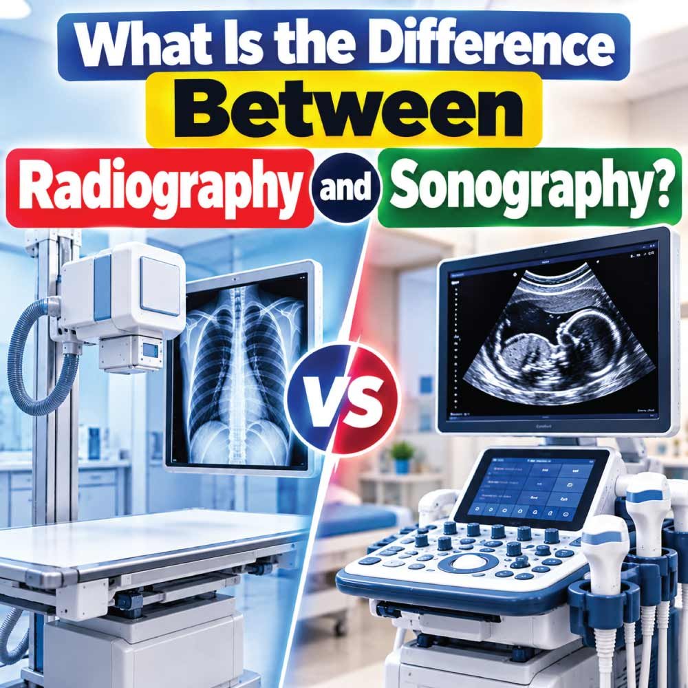
Radiography and sonography are both important medical imaging fields, but they use very different technologies to create images of the body. Understanding the difference between radiography and sonography is useful for patients comparing diagnostic tests, students exploring healthcare careers, and anyone interested in medical imaging.
Radiography primarily uses X-rays, a form of ionizing radiation, to produce images of internal structures. It is commonly associated with chest X-rays, bone imaging, dental imaging, and certain examinations performed with contrast material.
Sonography, also called ultrasound imaging, uses high-frequency sound waves rather than ionizing radiation. A sonographer moves a transducer over the body, and the returning sound waves are processed into images on a monitor.
The two professions also differ in equipment, examination techniques, training, working environments, and typical clinical applications. However, both require technical knowledge, attention to detail, communication skills, and an understanding of human anatomy.
The most important distinction is simple: radiography uses X-rays, while sonography uses sound waves.
How Radiography Works
Radiography creates images by directing X-rays through the body toward a detector. Different tissues absorb X-rays at different rates, allowing the resulting image to show variations in density.
For example, bones absorb considerably more X-rays than many surrounding soft tissues, which is why they appear prominently on conventional radiographic images.
During a typical examination, the radiographer positions the patient and the body part being examined. The equipment is then aligned to obtain the required projection before the exposure is made.
Modern radiography generally uses digital detectors rather than traditional photographic film. Digital images can be viewed, stored, transmitted, and evaluated through electronic medical imaging systems.
Radiography is particularly useful when the clinical question involves structures that are well demonstrated by X-ray imaging.
Common examples include:
- Fractures and other bone injuries
- Chest and lung examinations
- Certain joint and skeletal conditions
- Dental imaging
- Some abdominal examinations
- Imaging performed with contrast agents
- Specialized procedures using X-ray guidance
Radiography can also be part of more advanced imaging systems. Computed tomography, for example, uses X-rays to generate cross-sectional images, although CT is a distinct imaging modality rather than simply another name for conventional radiography.
How Sonography Works
Sonography uses high-frequency sound waves to produce images of internal body structures. The central piece of equipment is the ultrasound transducer, sometimes called an ultrasound probe.
The transducer sends sound waves into the body. When those waves encounter boundaries between different tissues, some of the sound is reflected back toward the probe.
The ultrasound system analyzes these returning echoes and converts them into an image.
Because ultrasound does not use ionizing radiation, it is widely used when real-time imaging is helpful and in situations where avoiding radiation exposure is important.
Sonography can be used to examine many different structures, including:
- The abdomen
- Gallbladder
- Liver
- Kidneys
- Thyroid
- Blood vessels
- Heart
- Reproductive organs
- Muscles and tendons
- Certain superficial soft tissues
Ultrasound can also show movement in real time. Doppler techniques allow clinicians to evaluate blood flow and are commonly used in vascular and cardiovascular examinations.
The quality of an ultrasound examination depends heavily on the operator’s technique. The sonographer may need to adjust the patient’s position, change the transducer angle, modify imaging settings, and obtain multiple views to answer the clinical question.
Radiography vs Sonography: Key Differences
Although both professions involve medical imaging, the underlying technology and examination process are substantially different.
| Feature | Radiography | Sonography |
|---|---|---|
| Main technology | X-rays | High-frequency sound waves |
| Ionizing radiation | Yes | No |
| Primary equipment | X-ray system and detector | Ultrasound system and transducer |
| Image production | X-ray absorption | Reflected sound-wave echoes |
| Real-time imaging | Limited in conventional radiography | Major strength |
| Operator involvement | Positioning and exposure technique | Continuous scanning and image optimization |
| Common applications | Bones, chest, some other structures | Soft tissues, organs, blood flow |
| Typical examination | X-ray | Ultrasound scan |
| Radiation precautions | Required | Not based on ionizing radiation |
| Dynamic assessment | Limited | Strong capability |
These differences do not mean one technology is universally better than the other. Each modality is designed to answer different clinical questions.
Radiography and Sonography Use Different Types of Energy
One of the easiest ways to understand the difference between radiography and sonography is to look at the energy each technology uses.
Radiography Uses X-Rays
X-rays are electromagnetic radiation. When X-rays pass through the body, tissues absorb different amounts of the radiation. A detector captures the transmitted X-rays and produces the radiographic image.
The amount of radiation used for an examination is controlled and optimized according to the procedure and patient circumstances. Radiation protection principles are an important part of radiography practice.
Sonography Uses Sound Waves
Ultrasound uses mechanical sound waves at frequencies above the range normally audible to humans.
The transducer both sends and receives sound waves. The system uses the returning echoes to determine the location and characteristics of structures within the scanned area.
Because the technologies work differently, the appearance of anatomical structures can also be very different between an X-ray image and an ultrasound image.
Which Examination Is Used for Different Conditions?
The choice between radiography and sonography depends on the clinical question, the body part being examined, and the type of information needed.
When Radiography May Be Used
Radiography is especially valuable for many bone and chest examinations.
For example, if a patient has suffered a suspected fracture, an X-ray may provide a rapid way to evaluate the affected bone.
Chest radiography is also widely used to examine the lungs, heart silhouette, and other structures visible on a chest image.
Radiographs can provide useful information about:
- Bone alignment
- Fractures
- Certain joint abnormalities
- Chest findings
- Some foreign objects
- Dental structures
When Sonography May Be Used
Ultrasound is particularly useful for many soft-tissue and fluid-related examinations.
For instance, abdominal ultrasound can help evaluate organs such as the liver, gallbladder, kidneys, and pancreas. Ultrasound can also distinguish certain fluid-filled structures from solid tissues.
Sonography is commonly used for:
- Abdominal imaging
- Obstetric examinations
- Pelvic imaging
- Vascular studies
- Thyroid examinations
- Musculoskeletal imaging
- Certain breast examinations
- Cardiac imaging through echocardiography
The appropriate examination is determined by the healthcare team based on the patient’s symptoms, medical history, and diagnostic needs.
Radiography vs Sonography for Bones and Soft Tissue
The type of tissue being investigated is an important factor when choosing an imaging method.
Radiography generally provides strong visualization of bones because dense mineralized tissue interacts strongly with X-rays. This makes conventional X-ray imaging a fundamental tool for many skeletal examinations.
Ultrasound, meanwhile, is particularly useful for evaluating soft tissues and structures that can be examined through an acoustic window.
For example, ultrasound can be useful when evaluating tendons and muscles because the operator can observe structures dynamically while the patient moves.
Neither method provides the same information in every situation.
A radiograph may demonstrate a fracture clearly, while ultrasound may provide more information about certain superficial soft-tissue structures. In some cases, healthcare professionals may use multiple imaging modalities because each provides complementary information.
Safety Differences Between Radiography and Sonography
Safety is another major difference between the two imaging technologies.
Radiography uses ionizing radiation. Medical imaging professionals therefore follow radiation protection principles designed to minimize unnecessary exposure while obtaining diagnostically useful images.
This does not mean that every X-ray examination is inherently unsafe. X-rays are widely used in healthcare because their diagnostic benefits can outweigh the associated radiation exposure when an examination is clinically appropriate.
Sonography does not use ionizing radiation. This makes it particularly useful when clinicians want real-time imaging without an X-ray exposure.
However, “no ionizing radiation” does not mean ultrasound should be treated as a completely unrestricted procedure. Ultrasound equipment produces acoustic energy, and examinations should be performed appropriately using established safety principles.
The key distinction is that the two modalities involve different forms of energy and therefore have different safety considerations.
What Does a Radiographer Do?
A radiographer, also called a radiologic technologist in some healthcare systems, is responsible for performing radiographic examinations and operating imaging equipment.
The job involves much more than simply pressing a button.
A radiographer may:
- Review the examination request
- Confirm patient identity and relevant information
- Explain the procedure
- Position the patient correctly
- Select appropriate technical factors
- Operate the X-ray equipment
- Apply radiation protection principles
- Check image quality
- Repeat an image when technically necessary
- Maintain accurate examination records
- Communicate with other healthcare professionals
Good positioning and technical accuracy are essential because an incorrectly positioned image may not answer the clinical question effectively.
Radiographers may work in hospitals, outpatient imaging centers, emergency departments, orthopedic settings, dental environments, or specialized radiology departments, depending on their qualifications.
Also Read:
What Does a Sonographer Do?
A sonographer performs ultrasound examinations using specialized equipment and transducers.
Sonography can be highly operator-dependent because the professional must actively manipulate the probe and interpret the appearance of anatomy while acquiring images.
Typical responsibilities can include:
- Reviewing the examination request
- Preparing and explaining the procedure
- Positioning the patient
- Selecting the appropriate transducer
- Applying ultrasound gel
- Scanning the required anatomy
- Adjusting imaging parameters
- Obtaining diagnostic images and measurements
- Evaluating anatomical structures during the examination
- Documenting relevant findings according to professional practice
In many clinical settings, the sonographer obtains and documents images for interpretation by an appropriately qualified physician or other authorized healthcare professional.
Sonographers may specialize in areas such as abdominal, vascular, cardiac, obstetric and gynecologic, breast, or musculoskeletal ultrasound.
Also Read:
Radiography and Sonography Career Comparison
Students considering medical imaging careers often compare radiography and sonography because both combine healthcare, technology, anatomy, and direct patient interaction.
The day-to-day experience, however, can be quite different.
| Career factor | Radiography | Sonography |
|---|---|---|
| Main imaging method | X-ray-based imaging | Ultrasound |
| Patient positioning | Important | Very important |
| Equipment operation | X-ray systems | Ultrasound systems |
| Image acquisition | Usually specific projections | Continuous scanning |
| Real-time image optimization | Limited | Central to the role |
| Radiation safety | Important | No ionizing radiation |
| Physical demands | Patient positioning and transfers | Repetitive scanning and positioning |
| Potential specialties | General, CT, mammography, interventional areas | Vascular, cardiac, abdominal, obstetric, musculoskeletal and others |
| Anatomy knowledge | Essential | Essential |
The exact scope of practice, education requirements, licensing, certification, and job titles vary by country and healthcare system.
Also Read:
Education and Training Requirements
Radiography and sonography both require structured education, but the specific pathway depends on where a person intends to practice.
Radiography programs typically cover subjects such as:
- Human anatomy
- Physiology
- Radiographic positioning
- Radiation physics
- Radiation protection
- Patient care
- Imaging procedures
- Clinical practice
Sonography education commonly includes:
- Anatomy
- Physiology
- Ultrasound physics
- Instrumentation
- Scanning techniques
- Pathophysiology
- Patient care
- Clinical training
Ultrasound physics is particularly important for sonographers because they need to understand how sound interacts with tissues and how technical settings affect image quality.
For both careers, classroom learning is only part of the preparation. Supervised clinical experience allows students to develop practical skills and learn how to communicate with patients in real healthcare environments.
Which Career Involves More Hands-On Imaging?
Both careers are hands-on, but the nature of the work differs.
In radiography, the professional typically positions the patient and equipment, selects the appropriate imaging approach, performs the exposure, and evaluates whether the resulting image meets technical requirements.
In sonography, the operator continuously controls the transducer and watches the image during scanning. Small changes in probe angle, pressure, position, or equipment settings can significantly affect what appears on the screen.
This makes manual scanning technique a central part of sonography.
For someone interested in continuous image acquisition and dynamic anatomy, the nature of ultrasound work may feel particularly hands-on. Someone interested in X-ray technology, positioning, and a wider pathway into radiologic imaging may find radiography more aligned with their interests.
These are descriptive differences rather than measures of which career is better.
Advantages and Limitations of Radiography
Radiography has several characteristics that make it valuable in modern healthcare.
Strengths of Radiography
- Fast acquisition for many examinations
- Strong visualization of many bones
- Widely available in healthcare settings
- Useful for chest imaging
- Can be performed with portable equipment in appropriate circumstances
- Forms part of more advanced X-ray-based imaging technologies
Limitations of Radiography
Radiographs are two-dimensional projections. Structures can overlap, which can sometimes make interpretation more difficult.
Soft tissues may also have less contrast on a conventional radiograph than on some other imaging modalities.
Additionally, because X-rays involve ionizing radiation, radiation protection must be incorporated into the examination process.
Advantages and Limitations of Sonography
Ultrasound also has a distinctive set of strengths and limitations.
Strengths of Sonography
- Does not use ionizing radiation
- Provides real-time imaging
- Useful for many soft-tissue examinations
- Can evaluate movement
- Doppler can assess blood flow
- Equipment can be relatively portable
- Can be useful for image-guided procedures
Limitations of Sonography
Ultrasound does not pass equally well through every type of tissue.
Air and bone can significantly limit visualization of structures located behind them. This means ultrasound may not be the ideal modality for every body region.
Image quality can also depend strongly on operator skill, patient anatomy, and the ability to obtain an appropriate acoustic window.
Body habitus and other technical factors can further affect the examination.
Can Radiography and Sonography Be Used Together?
Yes. Different imaging modalities can complement one another.
A patient may undergo an X-ray when the clinical question concerns a possible bone injury, while ultrasound may subsequently be used to investigate soft tissues or another structure.
In other situations, ultrasound may be used as an initial examination, followed by another imaging study if additional information is needed.
Medical imaging is not generally a competition between technologies. The purpose is to select the examination that can provide useful information for the particular clinical question.
The decision may depend on:
- The suspected condition
- The body part involved
- Patient age and circumstances
- Previous imaging
- Clinical symptoms
- Availability of equipment
- The information the healthcare professional needs
Radiographer vs Sonographer: Which Role Is Different?
The simplest distinction is that a radiographer primarily works with X-ray-based imaging, while a sonographer specializes in ultrasound examinations.
The workflow also differs.
A radiographer may perform a small number of carefully positioned exposures to create the required views. A sonographer typically spends more time actively scanning an area and adjusting the transducer throughout the examination.
Both professionals need strong anatomical knowledge and patient-care skills.
Both must also understand their equipment, follow professional standards, maintain accurate documentation, and recognize when image acquisition needs to be adjusted.
The biggest difference is therefore not the importance of the professions but the technology, imaging technique, and clinical applications involved.
Radiography and Sonography: Quick Summary
For readers looking for a straightforward answer, the comparison can be summarized as follows:
| Question | Radiography | Sonography |
|---|---|---|
| What energy is used? | X-rays | Sound waves |
| Does it use ionizing radiation? | Yes | No |
| What is the main device? | X-ray tube and detector | Ultrasound transducer |
| Best known for | Bones and chest imaging | Soft tissues and real-time imaging |
| Can it show movement? | Limited in conventional radiography | Yes |
| Can it assess blood flow? | Not its primary function | Yes, using Doppler |
| Is operator technique important? | Yes | Especially important |
| Common professional | Radiographer/radiologic technologist | Sonographer |
Frequently Asked Questions
Is sonography the same as radiography?
No. Sonography and radiography are different medical imaging techniques. Sonography uses high-frequency sound waves, while radiography uses X-rays.
Which uses radiation, radiography or sonography?
Radiography uses ionizing X-ray radiation. Sonography does not use ionizing radiation; it uses ultrasound sound waves.
Is an ultrasound better than an X-ray?
Neither is universally better. Each is suited to different clinical questions. X-rays are particularly useful for many bone and chest examinations, while ultrasound is valuable for many soft-tissue, fluid, vascular, and real-time imaging applications.
Do radiographers and sonographers do the same job?
No. Their responsibilities overlap in areas such as patient care, anatomy, image acquisition, and equipment operation, but they specialize in different imaging technologies.
Can ultrasound detect fractures?
Ultrasound can visualize some superficial structures and has applications in certain musculoskeletal assessments, but conventional X-ray remains an important imaging method for evaluating many suspected fractures.
Is sonography a medical imaging career?
Yes. Sonography is a healthcare imaging profession focused on obtaining diagnostic ultrasound examinations. Depending on the healthcare system, professionals may specialize in areas such as vascular, cardiac, abdominal, obstetric, gynecologic, or musculoskeletal sonography.
Final Takeaway: Radiography vs Sonography
The difference between radiography and sonography begins with the technology: radiography uses X-rays, whereas sonography uses high-frequency sound waves.
From there, the differences extend into image production, examination technique, equipment, safety considerations, clinical applications, and professional responsibilities.
Radiography is particularly established for skeletal and chest imaging, while sonography is especially valuable for many soft-tissue, vascular, fluid, and dynamic examinations.
For patients, the appropriate imaging test depends on the clinical question and the recommendation of the healthcare professional. For students, understanding these differences can help clarify what each medical imaging career involves.
Ultimately, radiography and sonography serve complementary purposes. Both depend on skilled professionals who understand anatomy, technology, patient care, and the importance of obtaining useful diagnostic images.

Zak is a dedicated medical and career writer specializing in sonography, healthcare education, and professional development. Through SonographerSalary.com, he shares in-depth insights on sonographer salaries, education pathways, and career tips to help readers build successful futures in medical imaging. His content combines accuracy with practical, easy-to-understand guidance, empowering students and professionals to make confident, informed career decisions.