
Sonography, also known as diagnostic medical ultrasound, is one of the most widely used imaging techniques in modern healthcare. If you have ever had an ultrasound during pregnancy, a scan for abdominal pain, or a heart examination called an echocardiogram, you’ve already experienced the power of sonography. What makes it unique is its use of high-frequency sound waves instead of radiation, which allows healthcare professionals to see inside the body safely and accurately.
This guide is designed to help you understand everything about sonography—what it is, how it works, what sonographers do, the training required, different specialties, salary expectations, and the benefits and limitations of ultrasound technology. Whether you are exploring a healthcare career, trying to understand a recent scan, or simply curious about medical imaging, this complete guide breaks it all down in an easy, friendly, and professional way.
What Is Sonography? A Clear and Simple Definition
Sonography is a medical imaging technique that uses sound waves to create real-time pictures of organs, tissues, blood flow, and developing fetuses. These images help doctors diagnose diseases, monitor medical conditions, and guide procedures. Unlike X-rays or CT scans, sonography does not expose patients to ionizing radiation, making it one of the safest imaging choices available.
At its core, the ultrasound transducer sends sound waves into the body, those waves bounce back, and a computer transforms the echoes into detailed images. This process happens instantly, offering live visuals that allow healthcare teams to watch body structures as they move.
How Sonography Works: The Science Made Simple
Understanding the technology behind ultrasound helps you appreciate its accuracy and value. The ultrasound machine uses a handheld device called a transducer, which emits sound waves that bounce off internal structures. The returning echoes help form images. This is similar to how bats use echolocation to navigate in the dark.
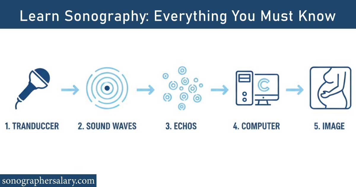
Here is a quick, integrated list of what happens during an ultrasound scan:
The transducer sends high-frequency sound waves into the body.
These waves bounce off organs, tissues, or fluid.
Echoes return to the transducer.
A computer processes them into images displayed on the screen.
What makes this process impressive is that it all happens in real time. Sonographers can capture movement as it occurs for example, watching the heart pump or tracking blood flow inside vessels.
Types of Sonography: A Look at the Main Specialties
Sonography includes multiple specialties because each area of the body requires a different scanning technique. Here are some of the most common branches explained in simple terms, with a helpful comparison chart below.
Specialties of Sonography
| Sonography Type | What It Examines | Common Use |
|---|---|---|
| Abdominal | Liver, gallbladder, kidneys, pancreas | Diagnose abdominal pain, organ problems |
| OB/GYN | Pregnancy, uterus, ovaries | Track fetal growth, diagnose women’s health issues |
| Cardiac (Echo) | Heart structure and function | Detect heart disease, monitor heart conditions |
| Vascular | Blood vessels, arteries, veins | Check circulation, diagnose blockages |
| MSK | Muscles, ligaments, joints | Sports injuries, tendon tears |
| Pediatric | Infants and children | Brain, abdomen, organs in newborns |
This variety makes sonography a flexible and rewarding career because you can specialize based on your interest.
📌 Diagnostic Imaging Courses Online offer a flexible and practical way to begin or advance your medical imaging career. This post breaks down course types, benefits, career paths, and real-world skills you’ll gain. Dive into the full guide to make the smartest choice and start your journey with confidence.
Why Sonography Is So Important in Modern Healthcare
Sonography plays a critical role in diagnosis and treatment. Because it is fast, affordable, and radiation-free, doctors rely on it for early detection of diseases. In emergency rooms, an ultrasound can help diagnose internal bleeding, appendicitis, or gallbladder issues within minutes. In cardiology, ultrasound helps evaluate heart failure and valve disorders. In obstetrics, it tracks fetal health throughout pregnancy.
Health systems prefer ultrasound because it offers the perfect blend of safety, accuracy, and convenience. That’s why millions of scans are performed worldwide every day.
What Sonographers Do: A Day in the Life
A diagnostic medical sonographer is the professional operating the ultrasound equipment. Their job is not simply taking pictures; it’s a mix of technical skill, patient care, and clinical judgment. Sonographers must understand anatomy, pathology, patient positioning, and proper scanning techniques.
They begin by reviewing a patient’s medical history and verifying the type of scan ordered. During the exam, they apply gel to improve sound contact, move the transducer around the target area, and capture detailed images from multiple angles. After scanning, they analyze the images, select the most relevant ones, and prepare them for the radiologist or physician.
Sonographers often work closely with cardiologists, OB/GYN specialists, surgeons, and emergency doctors, making teamwork an essential part of their role.
Real-Time Imaging: How Ultrasound Shows Movement Inside the Body
One of the most fascinating features of ultrasound is its ability to show movement. For example, in a vascular ultrasound, sonographers can watch blood flowing through arteries and veins. In an obstetric scan, they can see a fetus move, kick, or stretch. In cardiac sonography, the heart’s valves open and close right on the screen.
This dynamic imaging allows doctors to detect abnormalities instantly—such as irregular heartbeats, blocked arteries, or organ malfunctions. The real-time aspect makes ultrasound an indispensable tool for guiding procedures like biopsies or injections.
Here is a small reference table showing how real-time imaging benefits different specialties:
| Specialty | Real-Time Use | Example |
|---|---|---|
| Cardiac | Heart movement | Detect valve disease |
| OB/GYN | Fetal activity | Check fetal heartbeat |
| Vascular | Blood flow direction | Identify clots |
| MSK | Tendon movement | Diagnose tendon tears |
📌 High-Paying Medical Jobs Without a Degree can open life-changing opportunities for anyone who wants a stable, rewarding healthcare career. This post explains real roles, salaries, and how to start quickly. Read the full guide to discover which path fits you best and how you can begin earning sooner than you think.
Training Required: Education and Certification Paths
Becoming a sonographer requires specialized training. Most students enroll in an accredited ultrasound program lasting 1 to 4 years, depending on whether they choose a certificate, associate degree, or bachelor’s degree. Students learn anatomy, physics, imaging principles, and hands-on scanning.
Certification plays a major role in career growth. In the U.S., organizations like ARDMS, CCI, or ARRT offer recognized credentials. Many employers prefer or require certification because it assures high-quality images and safe practice.
Below is a simplified table showing common pathways:
| Education Level | Duration | Notes |
|---|---|---|
| Certificate | 12–18 months | For those already in healthcare |
| Associate Degree | 2 years | Most common entry route |
| Bachelor’s Degree | 4 years | Best for long-term career growth |
Sonography Salary Overview: Earnings and Career Growth
Sonographers are among the highest-paid allied health professionals without needing a medical degree. Salaries vary based on experience, location, and specialty. Cardiac and vascular sonographers typically earn more due to their specialized training.
Here is a salary overview chart:
| Experience Level | Estimated Yearly Salary |
|---|---|
| Entry-Level | $55,000–$70,000 |
| Mid-Level | $70,000–$85,000 |
| Senior-Level/Specialty | $90,000–$110,000+ |
High demand and advancing technology continue to increase job opportunities nationwide.
📌 Ultrasound Tech Programs provide a clear, practical path into a respected and growing healthcare career. This post explains program types, required skills, clinical training, and how to choose the right school. Read the full guide to understand the journey and start building your future in medical imaging with confidence.
Benefits of Sonography for Patients and Providers
Sonography offers many advantages that make it the first choice for evaluating countless medical conditions. It is safe, painless, and widely available. Patients do not need preparation for most scans and can return to normal activities immediately afterward. Doctors appreciate ultrasound because it is quick and cost-effective.
Below is a simple benefits comparison table:
| Benefit | Why It Matters |
|---|---|
| No radiation | Safe for all ages, including pregnancy |
| Affordable | Lower cost than CT/MRI |
| Fast results | Real-time imaging helps quick diagnosis |
| Widely available | Found in clinics, hospitals, ERs |
| Non-invasive | No needles or surgery required |
Common Mistakes People Make Before or During Ultrasound Exams
While ultrasound is usually simple, certain mistakes can affect image quality. One common issue is not following preparation instructions. For example, abdominal ultrasounds often require fasting so that gas in the stomach does not block sound waves. Another mistake is drinking too little water before a pelvic ultrasound, which requires a full bladder for clearer imaging.
Patients sometimes worry too much about the results or misinterpret what they see on the screen. It’s important to remember that only radiologists or physicians can make final diagnostic conclusions.
Communication between sonographers and patients is crucial. Asking questions and expressing discomfort ensures better accuracy and a more comfortable experience.
Limitations of Sonography: What Ultrasound Cannot Do
Although ultrasound is incredibly useful, it does have limitations. Sound waves cannot pass well through bone or air, which makes it less effective for imaging the lungs or certain areas of the brain. Extremely obese patients may also have reduced image quality because sound waves weaken when passing through thick tissue.
Below is a quick chart showing limitations:
| Limitation | Why It Occurs |
|---|---|
| Poor imaging through bone | Sound waves cannot penetrate solid bone |
| Poor imaging through gas | Air scatters sound waves |
| Reduced clarity in obesity | Sound waves weaken through thick tissue |
| Operator-dependent | Quality varies by skill level |
Even with these limitations, ultrasound remains one of the most reliable tools for soft-tissue imaging.
📌 Master Sonography Programs are designed for advanced learners aiming to excel in medical imaging and diagnostic expertise. This post covers program structure, specializations, career outcomes, and skills you’ll gain. Read the full guide to explore how a master’s degree can elevate your sonography career to the next level.
Future of Sonography: AI, Portable Devices, and Advanced Imaging
Sonography is rapidly evolving. Portable ultrasound machines now allow doctors to perform bedside scans, even in ambulances or remote areas. Artificial intelligence is improving image interpretation, helping detect abnormalities earlier and guiding less experienced operators.
3D and 4D ultrasound technologies create lifelike moving images, especially in obstetrics. Vascular and cardiac specialties now use advanced Doppler tools to evaluate blood flow with greater precision.
The future promises even more accurate, faster, and user-friendly ultrasound technology—making sonography an expanding field with endless potential.
Conclusion: Why Sonography Remains Essential in Healthcare
Sonography is one of the most important and versatile imaging tools in medicine today. It offers real-time, radiation-free imaging that helps diagnose diseases, guide treatments, and monitor health conditions. From pregnancy care to heart examinations, vascular studies, and emergency evaluations, ultrasound continues to improve patient outcomes across specialties.
For those exploring a healthcare career, sonography provides meaningful work, strong job security, high earning potential, and multiple paths for specialization. For patients, ultrasound ensures safe, fast, and accurate medical imaging right when it matters most.
Whether you are choosing a career or preparing for a scan, understanding sonography empowers you with confidence and clarity—making this essential technology even more valuable in your health journey.
📌 Ultrasound Tech: Complete Guide covers everything you need to know about becoming an ultrasound technician, from training and certifications to career opportunities and salaries. Read the full post to get expert insights, practical tips, and step-by-step guidance to launch a successful career in medical imaging.
FAQs:
What is sonography used for?
Sonography is used to create real-time images of organs, tissues, and blood flow without radiation. Doctors rely on this test to diagnose medical conditions early, guide procedures, monitor pregnancies, and evaluate pain or swelling. It’s a safe, noninvasive method that helps identify problems before they become serious.
Why do girls do sonography?
Girls often undergo sonography to check reproductive health, diagnose pelvic issues, or monitor pregnancy. It helps evaluate ovaries, uterus, menstrual-cycle concerns, and early pregnancy development. Doctors also use it to detect cysts, fibroids, or fertility problems. The test is safe, painless, and provides quick, detailed insights.
What is the difference between ultrasound and sonography?
Sonography is the process of performing an ultrasound scan, while ultrasound is the technology that uses sound waves to create images. In simple terms, ultrasound is the tool, and sonography is the technique. Both work together to visualize organs, track pregnancy growth, and diagnose medical conditions.
What is the highest paid sonographer?
The highest-paid sonography professionals are usually cardiac sonographers and vascular sonographers. They earn more because their work requires advanced training and highly detailed imaging skills. These specialists often work in hospitals and cardiovascular centers, where accurate scans are critical for diagnosing life-threatening heart and blood-vessel conditions.
How is a sonography test done?
A sonography test is done by applying warm gel on the skin and moving a handheld device called a transducer over the area. The device sends sound waves that create images on a screen. The procedure is painless, safe, and usually completed within 15–30 minutes depending on the exam type.
What is another term for sonography?
Another term for sonography is ultrasonography, which means using high-frequency sound waves to create internal body images. Some people also call it an ultrasound scan. All these names describe the same safe, radiation-free imaging technique commonly used for pregnancy, organ evaluation, and medical diagnosis.

Zak is a dedicated medical and career writer specializing in sonography, healthcare education, and professional development. Through SonographerSalary.com, he shares in-depth insights on sonographer salaries, education pathways, and career tips to help readers build successful futures in medical imaging. His content combines accuracy with practical, easy-to-understand guidance, empowering students and professionals to make confident, informed career decisions.