
Acoustic shadowing is an ultrasound artifact where a dark area appears behind a structure that strongly blocks or reflects sound waves, such as bone or a stone. It occurs because fewer echoes return to the transducer. This feature helps identify calcifications, gallstones, kidney stones, and other dense structures in medical imaging.
Ultrasound imaging is one of the most widely used diagnostic tools in modern medicine. It allows healthcare professionals to examine internal organs, tissues, blood vessels, and developing babies without exposing patients to radiation. During an ultrasound examination, sonographers and radiologists often encounter imaging phenomena known as artifacts. One of the most common and clinically important ultrasound artifacts is acoustic shadowing.
Acoustic shadowing can provide valuable diagnostic information and often helps doctors identify structures such as stones, calcifications, bones, and certain tumors. Understanding acoustic shadowing can help patients better interpret their ultrasound reports and appreciate how ultrasound images are analyzed.
In this complete guide, we will explain what acoustic shadowing is, how it occurs, its causes, types, clinical significance, and common examples seen in ultrasound imaging.
What Is Acoustic Shadowing?
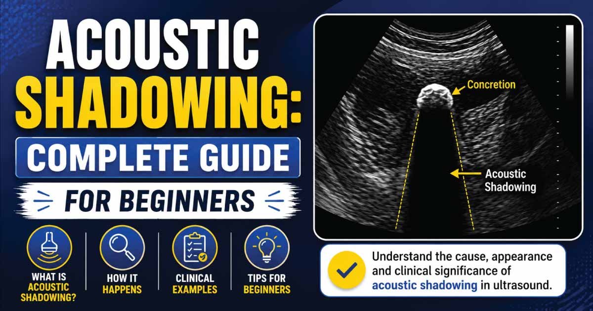
Acoustic shadowing is an ultrasound artifact that appears as a dark or black area behind a structure that strongly blocks or reflects ultrasound waves.
When ultrasound waves encounter a highly dense object, very little sound energy continues beyond that structure. As a result, the tissues located behind it receive fewer ultrasound waves and produce fewer echoes.
This creates a shadow-like appearance on the ultrasound image.
In simple terms, acoustic shadowing occurs when an object prevents ultrasound waves from reaching deeper tissues, creating a dark region behind it.
How Does Acoustic Shadowing Occur?
Ultrasound imaging works by sending high-frequency sound waves into the body.
These waves travel through tissues and return to the transducer after being reflected.
However, certain structures can:
- Absorb sound waves
- Reflect most sound waves
- Scatter sound waves
- Block sound transmission
When this happens, insufficient sound energy reaches deeper tissues.
The result is a dark shadow appearing behind the structure.
This dark area is known as an acoustic shadow.
How Does Acoustic Shadowing Appear on Ultrasound?
Acoustic shadowing usually appears as:
- A dark band
- A black streak
- A signal void
- An area lacking echoes
The shadow extends behind the structure responsible for blocking the ultrasound beam.
The size and intensity of the shadow depend on:
- Object density
- Object composition
- Ultrasound frequency
- Beam angle
- Depth of the structure
Why Is Acoustic Shadowing Important?
Although acoustic shadowing is technically an artifact, it often provides useful diagnostic information.
Doctors use acoustic shadowing to:
- Identify kidney stones
- Detect gallstones
- Recognize calcifications
- Evaluate bones
- Characterize tumors
- Confirm foreign bodies
In many cases, the presence of shadowing helps radiologists make a diagnosis more confidently.
Common Causes of Acoustic Shadowing
Several structures can produce acoustic shadowing.
Calcifications
Calcium strongly reflects ultrasound waves.
Examples include:
- Thyroid calcifications
- Breast calcifications
- Vascular calcifications
- Prostate calcifications
These often create distinct posterior acoustic shadows.
Gallstones
Gallstones are among the most classic causes of acoustic shadowing.
Because stones are dense, they block ultrasound waves and create a dark shadow behind them.
The combination of:
- Bright stone appearance
- Strong shadowing
helps confirm the diagnosis.
Kidney Stones
Renal stones also produce characteristic acoustic shadows.
The shadow helps distinguish stones from other bright structures within the kidney.
Bone
Bone surfaces strongly reflect ultrasound waves.
Examples include:
- Ribs
- Skull bones
- Long bones
These structures commonly generate prominent acoustic shadows.
Foreign Bodies
Certain foreign objects may cause shadowing.
Examples include:
- Metal fragments
- Surgical clips
- Glass pieces
- Certain implants
Shadowing helps locate and identify these materials.
Types of Acoustic Shadowing
Not all acoustic shadows look the same.
Different mechanisms can create different shadow patterns.
Clean Acoustic Shadowing
Clean shadowing appears as a well-defined dark area directly behind a dense structure.
Common causes include:
- Gallstones
- Kidney stones
- Bone
This type of shadow is often considered highly diagnostic.
Dirty Acoustic Shadowing
Dirty shadowing appears less distinct and often contains internal echoes.
It commonly occurs when ultrasound waves interact with gas.
Examples include:
- Bowel gas
- Air within tissues
- Lung interfaces
Dirty shadows often have a hazy appearance.
Partial Shadowing
Some structures only block part of the ultrasound beam.
This creates a less pronounced shadow.
Partial shadowing may occur with:
- Small calcifications
- Soft tissue masses
- Fibrotic tissue
Posterior Acoustic Shadowing
The most common form of shadowing is posterior acoustic shadowing.
The word “posterior” refers to the area located behind the structure relative to the ultrasound beam.
Posterior shadowing develops because:
- Sound waves are absorbed.
- Sound waves are reflected.
- Sound waves fail to penetrate deeper tissues.
This results in a dark region extending beyond the structure.
Acoustic Shadowing vs Posterior Enhancement
Acoustic shadowing is often confused with posterior acoustic enhancement.
However, they are opposite phenomena.
Acoustic Shadowing
Characteristics:
- Dark area behind structure
- Caused by sound attenuation
- Seen with stones and calcifications
Posterior Enhancement
Characteristics:
- Bright area behind structure
- Caused by increased sound transmission
- Seen behind fluid-filled cysts
Understanding the difference helps radiologists characterize lesions accurately.
Acoustic Shadowing in Gallstones
Gallstones are one of the most common examples of acoustic shadowing.
Typical ultrasound findings include:
- Bright echogenic stone
- Strong posterior shadow
- Mobile position within the gallbladder
The shadow confirms that the structure is likely a stone rather than sludge or debris.
Acoustic Shadowing in Kidney Stones
Kidney stones frequently produce:
- Bright echoes
- Posterior shadowing
- Twinkling artifact on Doppler imaging
Shadowing is especially useful when evaluating larger stones.
Smaller stones may produce weaker shadows.
Also Read:
Acoustic Shadowing in Thyroid Ultrasound
Thyroid nodules sometimes contain calcifications.
These calcifications can generate acoustic shadows.
The presence of shadowing may help identify:
- Benign calcified nodules
- Degenerating nodules
- Suspicious thyroid lesions
Radiologists evaluate shadowing along with other ultrasound features.
Acoustic Shadowing in Breast Ultrasound
Certain breast lesions create posterior acoustic shadowing.
Examples include:
- Fibroadenomas
- Scar tissue
- Calcified lesions
- Breast cancers
Although shadowing can be associated with malignancy, many benign lesions also produce shadows.
Additional imaging characteristics are always considered.
Acoustic Shadowing in Obstetric Ultrasound
During pregnancy, fetal bones create acoustic shadows.
Examples include:
- Skull
- Spine
- Long bones
These shadows are normal and expected.
However, excessive shadowing may occasionally limit visualization of underlying structures.
Acoustic Shadowing in Musculoskeletal Ultrasound
Musculoskeletal imaging frequently demonstrates acoustic shadowing.
Common sources include:
- Bones
- Calcified tendons
- Joint calcifications
- Orthopedic hardware
Shadowing can help identify abnormal calcium deposits within tendons and joints.
Also Read:
Advantages of Acoustic Shadowing
Acoustic shadowing is an ultrasound artifact that occurs when sound waves are blocked or strongly reflected by dense structures. Although it is an artifact, it provides important diagnostic clues that help clinicians identify and confirm various pathological conditions.
1. Improved Stone Detection
Acoustic shadowing is highly useful in detecting gallstones and kidney stones. These stones block ultrasound waves, creating a clear dark shadow behind them, which helps radiologists easily identify their presence and location with greater confidence.
2. Detection of Calcifications
Calcified tissues and lesions strongly reflect ultrasound waves, producing distinct shadowing. This makes it easier to recognize calcifications in organs or soft tissues, which may be associated with chronic disease, tumors, or degenerative changes.
3. Confirmation of Dense Structures
Shadowing helps confirm the presence of dense or highly reflective materials such as bone, stones, or foreign bodies. The presence of a clear acoustic shadow behind a structure supports its solid and calcified nature.
4. Enhanced Diagnostic Accuracy
Radiologists use acoustic shadowing as an important diagnostic clue during ultrasound interpretation. When combined with other imaging features, it improves diagnostic accuracy and helps differentiate between fluid-filled, solid, and calcified structures effectively.
Limitations of Acoustic Shadowing
Acoustic shadowing is a useful ultrasound artifact, but it also has several limitations. It can block important anatomical details, reduce image clarity, and sometimes make diagnosis difficult. Understanding these limitations is essential for accurate interpretation and reliable clinical assessment.
1. Obscures Deep Structures
Acoustic shadowing blocks the passage of ultrasound waves beyond dense objects like stones or bone. This prevents proper visualization of deeper tissues, making it difficult for clinicians to assess what lies behind the shadow and potentially missing important hidden abnormalities.
2. Reduced Image Quality
Large or strong shadows can significantly reduce overall image clarity. When sound waves are fully reflected or absorbed, the area behind the structure appears dark, which limits diagnostic detail and can interfere with accurate interpretation of surrounding anatomy.
3. Potential Diagnostic Challenges
Acoustic shadowing may hide lesions or abnormalities located deeper in the body. This can create diagnostic uncertainty, especially when the shadow overlaps clinically important regions, requiring additional imaging methods to confirm or rule out underlying conditions effectively.
4. Operator Dependence
Accurate interpretation of acoustic shadowing depends heavily on the skill and experience of the ultrasound operator. Poor technique, incorrect probe positioning, or suboptimal settings may exaggerate or misrepresent shadowing, leading to incomplete or misleading diagnostic information.
How Do Sonographers Reduce Unwanted Shadowing?
Sonographers use several techniques to improve image quality.
These include:
- Changing probe angle
- Adjusting frequency
- Using multiple scanning planes
- Repositioning the patient
- Applying additional imaging modes
These methods help evaluate structures hidden behind shadows.
Can Acoustic Shadowing Indicate Cancer?
Not necessarily.
Acoustic shadowing itself is not a diagnosis.
While some cancers may produce shadowing, many benign conditions do as well.
Examples of benign causes include:
- Gallstones
- Kidney stones
- Scar tissue
- Benign calcifications
Doctors always evaluate:
- Shape
- Size
- Margins
- Blood flow
- Clinical history
before determining whether a lesion is suspicious.
Common Ultrasound Terms Related to Acoustic Shadowing
You may encounter these related ultrasound terms:
- Echogenicity
- Echogenic
- Hypoechoic
- Hyperechoic
- Anechoic
- Isoechoic
- Posterior Enhancement
- Reverberation Artifact
- Edge Shadowing
- Attenuation
Understanding these concepts helps make ultrasound reports easier to interpret.
Also Read:
When Should You Discuss Acoustic Shadowing with Your Doctor?
If your ultrasound report mentions acoustic shadowing, you should discuss the finding with your healthcare provider.
Questions to ask may include:
- What structure is causing the shadow?
- Is the finding normal?
- Does it suggest a stone or calcification?
- Is further testing required?
- Should follow-up imaging be performed?
Your doctor can explain the significance based on your specific medical situation.
Conclusion
Acoustic shadowing is a common ultrasound artifact that occurs when a dense structure blocks or reflects ultrasound waves, creating a dark shadow behind it. Although it is technically an imaging artifact, it often provides valuable diagnostic information and helps identify stones, calcifications, bones, foreign bodies, and certain tissue abnormalities.
The presence of acoustic shadowing does not automatically indicate a serious medical problem. In many cases, it helps radiologists confirm normal anatomical structures or diagnose common conditions such as gallstones and kidney stones. Understanding acoustic shadowing can make ultrasound reports easier to interpret and improve communication between patients and healthcare providers.
If your ultrasound report mentions acoustic shadowing, consult your healthcare provider for an accurate explanation of its significance and whether any further evaluation is needed.

Zak is a dedicated medical and career writer specializing in sonography, healthcare education, and professional development. Through SonographerSalary.com, he shares in-depth insights on sonographer salaries, education pathways, and career tips to help readers build successful futures in medical imaging. His content combines accuracy with practical, easy-to-understand guidance, empowering students and professionals to make confident, informed career decisions.