
Posterior enhancement is an ultrasound artifact where tissues behind a fluid-filled or low-attenuation structure appear brighter than surrounding areas. It occurs because sound waves pass easily through fluid and gain increased strength beyond it. This feature helps identify cysts, fluid collections, and vascular structures during ultrasound examinations for accurate diagnosis.
Ultrasound imaging is one of the most valuable diagnostic tools used in modern medicine. It helps healthcare professionals examine internal organs, soft tissues, blood vessels, and developing babies without exposing patients to radiation. During an ultrasound examination, radiologists and sonographers often observe imaging phenomena known as ultrasound artifacts. One of the most common and clinically useful artifacts is posterior enhancement.
Posterior enhancement provides important diagnostic clues and helps healthcare providers identify fluid-filled structures such as cysts, the urinary bladder, gallbladder, and certain types of masses. Understanding posterior enhancement can help patients better interpret ultrasound reports and understand the significance of their imaging findings.
In this complete guide, we will explain what posterior enhancement is, how it occurs, its causes, clinical importance, and common examples seen in ultrasound imaging.
What Is Posterior Enhancement?
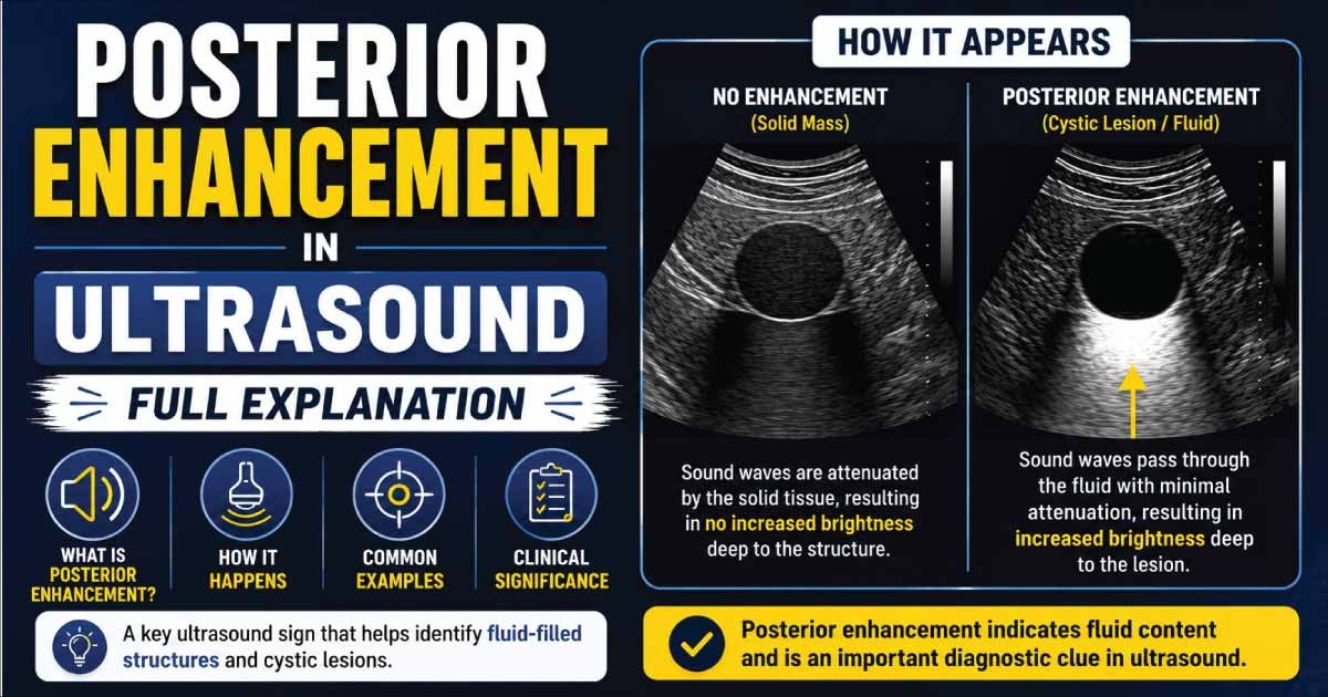
Posterior enhancement, also known as posterior acoustic enhancement or through transmission, is an ultrasound artifact that appears as an unusually bright area behind a structure that allows ultrasound waves to pass through easily.
When ultrasound waves travel through a fluid-filled structure, very little sound energy is absorbed or reflected. As a result, more ultrasound energy reaches the tissues located behind the structure.
These deeper tissues return stronger echoes, causing them to appear brighter than expected on the ultrasound image.
In simple terms, posterior enhancement occurs when a structure transmits sound waves efficiently, making the tissues behind it appear brighter.
How Does Posterior Enhancement Occur?
Ultrasound imaging works by sending sound waves into the body.
As the sound waves travel through tissues, some are reflected back while others continue deeper into the body.
Certain structures, particularly those containing fluid, cause very little attenuation of ultrasound waves.
Because minimal energy is lost:
- More sound waves pass through the structure.
- More energy reaches deeper tissues.
- Stronger echoes return from those tissues.
- The area behind the structure appears brighter.
This increased brightness is called posterior enhancement.
How Does Posterior Enhancement Appear on Ultrasound?
Posterior enhancement appears as:
- A bright area behind a structure
- Increased echogenicity in deeper tissues
- A white or lighter region extending beyond a lesion
- Improved visualization of tissues located behind fluid
The enhancement follows the path of the ultrasound beam and is most noticeable directly behind the structure responsible for the artifact.
Why Is Posterior Enhancement Important?
Posterior enhancement is one of the most useful ultrasound artifacts because it helps radiologists identify the nature of a lesion.
It can help:
- Differentiate cysts from solid masses
- Identify fluid-filled structures
- Improve lesion characterization
- Increase diagnostic confidence
- Guide treatment decisions
The presence of posterior enhancement often suggests that a structure contains fluid or material that transmits ultrasound efficiently.
Common Causes of Posterior Enhancement
Several structures can produce posterior enhancement.
Simple Cysts
Simple cysts are the classic example.
Because cysts are filled with fluid, ultrasound waves pass through them easily, producing strong posterior enhancement.
Examples include:
- Kidney cysts
- Liver cysts
- Ovarian cysts
- Breast cysts
Urinary Bladder
The bladder contains urine, which allows sound waves to travel with minimal attenuation.
This frequently results in prominent posterior enhancement.
Gallbladder
The gallbladder contains bile and commonly demonstrates posterior enhancement.
This finding helps distinguish fluid-filled structures from solid tissues.
Fluid Collections
Various fluid collections may produce enhancement.
Examples include:
- Seromas
- Hematomas
- Abscesses
- Pleural effusions
Certain Soft Tissue Lesions
Some benign and malignant lesions contain fluid components that generate posterior enhancement.
Posterior Enhancement vs Acoustic Shadowing
Posterior enhancement is often confused with acoustic shadowing.
However, these artifacts are opposites.
Posterior Enhancement
Characteristics:
- Bright area behind structure
- Increased sound transmission
- Usually associated with fluid-filled structures
Acoustic Shadowing
Characteristics:
- Dark area behind structure
- Reduced sound transmission
- Usually associated with stones, bone, or calcifications
Understanding this difference is essential for accurate ultrasound interpretation.
Posterior Enhancement in Simple Cysts
Simple cysts are one of the most common causes of posterior enhancement.
Typical ultrasound features include:
- Anechoic appearance
- Thin walls
- Smooth borders
- Strong posterior enhancement
These findings often allow radiologists to confidently diagnose a benign cyst.
Posterior Enhancement in Breast Ultrasound
Breast ultrasound frequently uses posterior enhancement to characterize lesions.
Breast cysts commonly demonstrate:
- Anechoic contents
- Smooth margins
- Posterior enhancement
Certain solid breast lesions may also produce enhancement, which is why radiologists evaluate multiple features before making a diagnosis.
Posterior Enhancement in Liver Ultrasound
Liver cysts often exhibit strong posterior enhancement.
Other liver lesions that may show enhancement include:
- Simple hepatic cysts
- Complex cysts
- Some vascular lesions
The enhancement pattern helps radiologists narrow the differential diagnosis.
Posterior Enhancement in Kidney Ultrasound
Kidney imaging commonly demonstrates posterior enhancement behind renal cysts.
Typical findings include:
- Round fluid-filled lesion
- No internal echoes
- Bright area behind the cyst
These characteristics often indicate a simple benign renal cyst.
Posterior Enhancement in Ovarian Ultrasound
Ovarian cysts frequently produce posterior enhancement.
Examples include:
- Functional cysts
- Follicular cysts
- Corpus luteum cysts
The enhancement helps confirm the presence of fluid within the lesion.
Posterior Enhancement in Thyroid Ultrasound
Some thyroid nodules contain fluid components.
These nodules may demonstrate posterior enhancement, particularly when they are:
- Predominantly cystic
- Colloid-filled
- Benign degenerative nodules
Radiologists evaluate enhancement alongside other ultrasound characteristics.
Posterior Enhancement in Obstetric Ultrasound
During pregnancy, fluid-filled structures naturally create enhancement.
Examples include:
- Amniotic fluid
- Fetal bladder
- Certain fetal cysts
These findings are often normal and expected.
How Radiologists Use Posterior Enhancement
Posterior enhancement helps healthcare providers determine whether a lesion is likely:
- Fluid-filled
- Solid
- Mixed solid and cystic
It is one of the key criteria used when evaluating masses throughout the body.
Radiologists combine enhancement findings with:
- Shape
- Size
- Borders
- Internal echoes
- Blood flow patterns
- Patient history
to reach a diagnosis.
Also Read:
Advantages of Posterior Enhancement
Posterior enhancement is an important ultrasound phenomenon that provides valuable diagnostic information, especially in fluid-filled structures. It helps clinicians interpret tissue characteristics more accurately and improves overall image analysis.
1. Improves Cyst Detection
Posterior enhancement makes fluid-filled lesions such as cysts appear brighter behind their walls. This clear enhancement helps radiologists easily identify cysts and distinguish them from solid masses during ultrasound evaluation.
2. Enhances Diagnostic Accuracy
This phenomenon provides additional clues about tissue composition. By observing how sound waves behave behind a structure, radiologists can better understand whether a lesion is fluid-filled, solid, or complex, improving overall diagnostic confidence.
3. Helps Differentiate Lesions
Posterior enhancement is especially useful in distinguishing cystic lesions from solid tumors. Cysts typically show strong enhancement, while solid masses do not, making this feature an important factor in lesion characterization.
4. Improves Visualization
Posterior enhancement increases visibility of deeper tissues located behind fluid-filled structures. Since sound waves travel easily through fluid, the area beyond becomes clearer, allowing better evaluation of surrounding anatomy and hidden structures.
Limitations of Posterior Enhancement
Posterior enhancement is a useful ultrasound artifact, but it has important limitations in diagnosis. It is not specific to one disease type and can appear in both benign and malignant conditions. Proper interpretation always requires correlation with clinical findings and other imaging features.
1. Not Specific for Benign Disease
Posterior enhancement is commonly seen in cystic or fluid-filled structures, but it is not limited to benign lesions. Some malignant or complex masses may also show this feature, so it cannot be used alone to confirm a harmless condition.
2. Can Mimic Pathology
Strong posterior enhancement may create areas of increased brightness behind a structure, which can sometimes be misleading. This effect may obscure true abnormalities or create false impressions, making accurate interpretation more challenging for clinicians during ultrasound evaluation.
3. Requires Experienced Evaluation
Correct assessment of posterior enhancement depends heavily on the skill of the radiologist or sonographer. It must always be interpreted along with lesion shape, margins, vascularity, and patient history to avoid diagnostic errors and improve accuracy.
4. May Occur in Multiple Conditions
Posterior enhancement can be seen in many different conditions, including cysts, abscesses, and some tumors. Because multiple diseases share this feature, it cannot be used as a standalone diagnostic marker and requires full clinical correlation.
Can Posterior Enhancement Indicate Cancer?
Posterior enhancement alone does not indicate cancer.
Many completely benign conditions produce strong enhancement.
Examples include:
- Simple cysts
- Functional ovarian cysts
- Benign fluid collections
- Normal bladder contents
Some cancers may contain fluid-filled components and also show enhancement.
For this reason, doctors evaluate additional imaging features before determining whether a lesion is suspicious.
How Is Posterior Enhancement Evaluated?
Posterior enhancement is evaluated by combining ultrasound imaging features with Doppler findings and patient clinical information. Doctors carefully analyze lesion characteristics, internal structure, and surrounding tissue changes to determine whether the enhancement is associated with benign or more complex pathology.
1. Lesion Shape
The shape of the lesion helps in initial assessment. Round or oval lesions are more commonly associated with benign cystic structures, while irregular shapes may raise suspicion and require further evaluation.
2. Wall Characteristics
Wall thickness and appearance are important indicators. Thin, smooth walls often suggest simple cysts, whereas thick or irregular walls may indicate infection, hemorrhage, or potentially malignant lesions needing closer investigation.
3. Internal Echoes
Internal echo patterns help define lesion content. Pure fluid appears anechoic and typically shows strong posterior enhancement, while internal echoes may suggest debris, blood, or solid components within a complex lesion.
4. Blood Flow
Doppler ultrasound is used to assess vascularity within or around the lesion. Absence of blood flow usually supports a simple cyst, while increased or abnormal vascularity may indicate inflammation or neoplastic processes.
5. Associated Findings
Doctors also evaluate nearby structures for additional abnormalities. The presence of edema, calcification, or adjacent tissue changes can provide important diagnostic clues and help narrow down the possible underlying condition.
6. Patient Symptoms
Clinical symptoms such as pain, fever, or swelling are always considered. Imaging findings are interpreted alongside patient history to ensure an accurate and complete diagnosis, improving confidence in clinical decision-making.
Also Read:
Common Ultrasound Terms Related to Posterior Enhancement
You may encounter several related ultrasound terms, including:
- Echogenicity
- Anechoic
- Hypoechoic
- Hyperechoic
- Isoechoic
- Acoustic Shadowing
- Through Transmission
- Attenuation
- Doppler Flow
- Reverberation Artifact
Understanding these terms can make ultrasound reports easier to interpret.
When Should You Discuss Posterior Enhancement with Your Doctor?
If your ultrasound report mentions posterior enhancement, consider asking:
- What structure is causing the enhancement?
- Does it represent a cyst?
- Is the finding benign?
- Are additional tests required?
- Should follow-up imaging be performed?
Your healthcare provider can explain the significance of the finding based on your medical history and imaging results.
Conclusion
Posterior enhancement is a common and highly useful ultrasound artifact that occurs when sound waves pass easily through a structure, allowing more energy to reach deeper tissues. This creates a brighter area behind the structure on the ultrasound image.
The finding is most commonly associated with fluid-filled structures such as cysts, the urinary bladder, gallbladder, and various fluid collections. Posterior enhancement often helps radiologists differentiate cystic lesions from solid masses and plays an important role in ultrasound diagnosis.
Although posterior enhancement is generally associated with benign fluid-filled structures, it should always be interpreted alongside other imaging features and clinical information. If your ultrasound report mentions posterior enhancement, discussing the results with your healthcare provider will help ensure an accurate understanding of the finding and any necessary follow-up care.
Also Read:

Zak is a dedicated medical and career writer specializing in sonography, healthcare education, and professional development. Through SonographerSalary.com, he shares in-depth insights on sonographer salaries, education pathways, and career tips to help readers build successful futures in medical imaging. His content combines accuracy with practical, easy-to-understand guidance, empowering students and professionals to make confident, informed career decisions.