
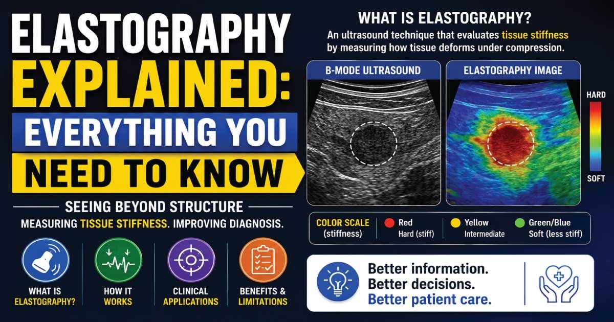
Elastography is an advanced ultrasound technique that measures tissue stiffness by assessing how tissues deform under pressure. Hard tissues, such as tumors or fibrosis, appear differently from soft tissues. It helps in early detection and characterization of liver disease, breast lesions, and other conditions, improving diagnostic accuracy and clinical decision-making.
Medical imaging has advanced significantly over the past few decades, allowing healthcare professionals to diagnose diseases earlier and more accurately than ever before. One of the most innovative developments in ultrasound imaging is Elastography, a technique that helps evaluate the stiffness or elasticity of tissues inside the body.
Traditional ultrasound provides information about the size, shape, and appearance of organs and lesions. Elastography adds another layer of information by measuring tissue stiffness, which can help distinguish between healthy and diseased tissues. This technology is widely used in liver imaging, breast examinations, thyroid evaluations, and many other medical applications.
In this complete guide, you will learn what elastography is, how it works, its types, benefits, limitations, clinical applications, and why it has become an important tool in modern diagnostic imaging.
What Is Elastography?
Elastography is a specialized imaging technique that measures the stiffness or elasticity of tissues.
The word “elastography” comes from:
- Elasticity = the ability of tissue to stretch and return to its original shape.
- Graphy = imaging or recording.
In simple terms, elastography helps doctors determine whether tissue is soft or stiff.
Healthy tissues are often more flexible, while diseased tissues may become harder due to inflammation, fibrosis, scarring, or cancer.
By measuring tissue stiffness, elastography provides valuable diagnostic information that may not be visible on conventional ultrasound.
Why Is Tissue Stiffness Important?
Many diseases alter the mechanical properties of tissues.
For example:
- Liver fibrosis causes the liver to become stiffer.
- Certain cancers are harder than surrounding tissues.
- Scar tissue is often less elastic than normal tissue.
- Chronic inflammation may increase tissue stiffness.
Measuring stiffness can help doctors:
- Detect disease earlier.
- Monitor disease progression.
- Evaluate treatment effectiveness.
- Reduce the need for invasive procedures.
How Does Elastography Work?
Elastography works by applying mechanical force or vibrations to tissues and measuring how they respond.
Soft tissues deform more easily.
Stiff tissues resist deformation.
The ultrasound machine analyzes tissue movement and creates an image or numerical measurement that reflects tissue stiffness.
The results may be displayed as:
- Color maps
- Quantitative measurements
- Elasticity scores
- Stiffness values
This information helps healthcare providers evaluate tissue health.
The Basic Principle of Elastography
The concept behind elastography is similar to what doctors do during a physical examination.
For example:
- A physician may press on an area of the body.
- Hard lumps feel different from soft tissue.
- Elastography performs a similar assessment using ultrasound technology.
Instead of relying on touch alone, the ultrasound system measures tissue stiffness objectively and accurately.
Types of Elastography
Several elastography techniques are used in modern medical imaging.
Strain Elastography
Strain elastography evaluates tissue deformation after slight compression.
The ultrasound probe applies gentle pressure, and the machine measures how much the tissue compresses.
Characteristics:
- Qualitative assessment
- Color-coded images
- Commonly used in breast and thyroid imaging
Soft tissues compress more easily than stiff tissues.
Shear Wave Elastography
Shear Wave Elastography (SWE) is one of the most advanced forms of elastography.
It generates mechanical waves within tissues and measures their speed.
Key principle:
- Faster wave speed = stiffer tissue
- Slower wave speed = softer tissue
Advantages include:
- Quantitative measurements
- High reproducibility
- Improved diagnostic accuracy
Transient Elastography
Transient Elastography is commonly used to assess liver stiffness.
A mechanical pulse creates a shear wave, and the system measures its propagation through the liver.
One of the best-known systems uses this technology for liver fibrosis assessment.
Point Shear Wave Elastography
This technique measures stiffness at a specific location within tissue.
It provides:
- Numerical values
- Targeted assessment
- Accurate focal measurements
How Are Elastography Results Displayed?
Results may be presented in different ways depending on the system.
Color Elastograms
Different colors represent different stiffness levels.
Typically:
- Blue indicates softer tissue.
- Red indicates stiffer tissue.
- Green and yellow represent intermediate stiffness.
Color scales may vary between manufacturers.
Numerical Measurements
Some systems provide quantitative values measured in:
- Kilopascals (kPa)
- Meters per second (m/s)
These measurements allow objective comparison over time.
Elastography of the Liver
Liver imaging is the most common application of elastography.
Why Liver Elastography Is Important
Chronic liver diseases often cause fibrosis.
Fibrosis gradually increases liver stiffness.
Elastography helps assess:
- Liver fibrosis
- Cirrhosis
- Chronic hepatitis
- Fatty liver disease
Advantages Over Liver Biopsy
Historically, liver biopsy was the standard method for evaluating fibrosis.
Elastography offers several advantages:
- Non-invasive
- Faster
- Safer
- Repeatable
- Less expensive
For many patients, elastography reduces the need for biopsy.
Breast Elastography
Breast elastography helps evaluate breast masses.
Cancerous tumors are often stiffer than surrounding tissue.
Benefits include:
- Better lesion characterization
- Reduced unnecessary biopsies
- Improved diagnostic confidence
Doctors often use elastography alongside conventional breast ultrasound.
Thyroid Elastography
Thyroid nodules are extremely common.
Most nodules are benign, but some require further investigation.
Elastography helps determine:
- Nodule stiffness
- Cancer risk assessment
- Need for biopsy
Stiffer nodules may require additional evaluation.
Prostate Elastography
Prostate elastography assists in detecting areas of abnormal tissue stiffness.
Potential applications include:
- Prostate cancer evaluation
- Targeted biopsy guidance
- Monitoring disease progression
Research continues to expand its clinical role.
Pancreatic Elastography
The pancreas can also be assessed using elastography.
Potential uses include:
- Chronic pancreatitis evaluation
- Pancreatic tumor assessment
- Fibrosis detection
This area continues to develop as technology improves.
Musculoskeletal Elastography
Elastography is increasingly used in sports medicine and musculoskeletal imaging.
Applications include:
- Tendon injuries
- Muscle disorders
- Ligament evaluation
- Scar tissue assessment
It helps clinicians better understand tissue healing and injury severity.
Elastography in Oncology
Cancer diagnosis is one of the most promising uses of elastography.
Many malignant tumors exhibit increased stiffness compared to normal tissue.
Elastography may assist with:
- Tumor detection
- Tumor characterization
- Treatment monitoring
- Biopsy planning
However, stiffness alone cannot confirm cancer.
Also Read:
Advantages of Elastography
Elastography is an advanced ultrasound technique that measures tissue stiffness to help identify and evaluate abnormalities. It provides additional functional information beyond standard imaging, improving diagnostic confidence and reducing the need for invasive procedures in many cases.
1. Non-Invasive
Elastography is completely non-invasive, meaning no surgery, needles, or incisions are required. This makes it a safe and comfortable method for evaluating tissue characteristics in various organs.
2. Painless
Most elastography examinations are painless and well tolerated by patients. The procedure is similar to a routine ultrasound scan and does not usually cause discomfort.
3. Quick Procedure
Elastography studies are typically completed within a few minutes. This makes it an efficient diagnostic tool that can easily be integrated into standard ultrasound examinations without significantly increasing exam time.
4. Real-Time Results
The technique provides immediate imaging and stiffness measurements during the scan. This allows clinicians to quickly assess tissue properties and make faster clinical decisions.
5. Improved Diagnostic Information
By measuring tissue stiffness, elastography adds valuable diagnostic information that is not available in conventional ultrasound. This helps in better characterization of lesions and improves overall diagnostic accuracy.
6. Reduced Need for Biopsy
In some cases, elastography can help differentiate benign from suspicious lesions, potentially reducing the need for invasive biopsy procedures. This improves patient comfort and reduces procedural risks.
Limitations of Elastography
Although elastography is a powerful diagnostic tool, it has certain limitations that can affect accuracy and interpretation. These factors must always be considered when evaluating results.
1. Operator Dependence
The accuracy of elastography results can vary depending on the skill and technique of the operator. Proper probe placement and correct settings are essential for reliable measurements.
2. Patient Factors
Patient body habitus, especially obesity or thick subcutaneous tissue, can reduce image quality. These factors may make it more difficult to obtain accurate stiffness measurements.
3. Motion Artifacts
Patient movement, breathing, or involuntary motion can affect elastography readings. These artifacts may lead to variability in results or reduced image clarity.
4. Not a Standalone Diagnostic Tool
Elastography should not be used alone for diagnosis. It must always be interpreted along with conventional ultrasound findings, clinical history, and other diagnostic tests for accurate assessment.
5. Equipment Variability
Different ultrasound systems and manufacturers may use different elastography techniques and measurement scales. This variability can affect consistency and comparison of results across different machines.
Also Read:
Who May Need Elastography?
Your doctor may recommend elastography if you have:
- Chronic liver disease
- Hepatitis
- Fatty liver disease
- Breast lumps
- Thyroid nodules
- Suspected fibrosis
- Certain cancers
- Abnormal imaging findings
The examination helps provide additional diagnostic information.
What Happens During an Elastography Exam?
An elastography exam is a simple, non-invasive imaging procedure that is performed in a similar way to a standard ultrasound scan. It helps evaluate tissue stiffness in real time, providing additional diagnostic information without causing discomfort to the patient.
1. Preparation
At the beginning of the exam, the patient is usually asked to lie down on an examination table in a comfortable position. The area being examined is exposed so that proper imaging can be performed.
2. Gel Application
A water-based ultrasound gel is applied to the skin over the target area. This gel helps improve sound wave transmission between the transducer and the body, ensuring clearer and more accurate images.
3. Probe Placement
The sonographer places the ultrasound transducer on the gel-covered area. Gentle pressure is applied to obtain optimal contact and stable imaging of the region of interest for elastography measurements.
4. Image Acquisition
The ultrasound machine captures elastography data by measuring tissue stiffness and generating real-time images or color maps. These measurements help differentiate between soft and stiff tissues within the examined area.
5. Interpretation
After the scan, a radiologist or physician reviews the elastography results. They analyze tissue stiffness patterns along with standard ultrasound images to reach an accurate diagnostic conclusion.
Most elastography examinations are completed within 10 to 30 minutes, depending on the area being studied and the complexity of the case.
Also Read:
Is Elastography Safe?
Yes.
Elastography uses ultrasound waves, which do not involve ionizing radiation.
It is considered:
- Safe
- Non-invasive
- Radiation-free
- Well tolerated
The procedure is widely used in both adults and children.
Common Terms Related to Elastography
When learning about elastography, you may encounter related terms such as:
- Tissue Stiffness
- Elasticity
- Fibrosis
- Shear Wave Elastography
- Strain Elastography
- Transient Elastography
- Liver Fibrosis
- Ultrasound Imaging
- Biopsy
- Quantitative Imaging
Understanding these terms can make elastography reports easier to interpret.
The Future of Elastography
Elastography continues to evolve rapidly.
Future advancements may include:
- Higher-resolution imaging
- Artificial intelligence integration
- Improved cancer detection
- Enhanced liver disease monitoring
- More precise quantitative measurements
These innovations are expected to further improve patient care and diagnostic accuracy.
Conclusion
Elastography is an advanced ultrasound imaging technique that measures tissue stiffness and elasticity. By providing information beyond traditional ultrasound images, it helps healthcare professionals evaluate fibrosis, inflammation, tumors, and other tissue abnormalities.
Today, elastography is widely used in liver, breast, thyroid, prostate, pancreatic, and musculoskeletal imaging. Its ability to provide non-invasive, real-time assessments of tissue health has made it one of the most valuable innovations in modern diagnostic ultrasound.
As technology continues to improve, elastography will likely play an even greater role in disease detection, treatment monitoring, and personalized patient care.

Zak is a dedicated medical and career writer specializing in sonography, healthcare education, and professional development. Through SonographerSalary.com, he shares in-depth insights on sonographer salaries, education pathways, and career tips to help readers build successful futures in medical imaging. His content combines accuracy with practical, easy-to-understand guidance, empowering students and professionals to make confident, informed career decisions.