
Echocardiography is one of the most important imaging techniques used in modern cardiology. It provides real-time images of the heart using ultrasound waves, allowing doctors to evaluate heart structure, function, blood flow, and overall cardiovascular health without surgery or radiation exposure.
Millions of echocardiograms are performed every year to diagnose and monitor heart diseases. Whether a patient has chest pain, shortness of breath, irregular heartbeat, heart murmurs, or suspected heart valve disease, echocardiography often serves as the first-line diagnostic tool.
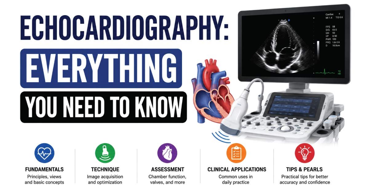
This comprehensive guide explains what echocardiography is, how it works, its types, uses, benefits, limitations, and what patients can expect during the procedure.
What Is Echocardiography?
Echocardiography is a medical imaging procedure that uses high-frequency sound waves (ultrasound) to create moving pictures of the heart.
The test allows healthcare providers to visualize:
- Heart chambers
- Heart valves
- Heart walls
- Blood vessels connected to the heart
- Blood flow through the heart
- Heart pumping function
Because ultrasound does not use ionizing radiation, echocardiography is considered a safe and non-invasive imaging method.
An echocardiogram is often referred to as an “echo.”
How Does Echocardiography Work?
Echocardiography works on the principle of ultrasound imaging.
A device called a transducer emits high-frequency sound waves into the body. These sound waves bounce off heart structures and return to the transducer as echoes.
The ultrasound machine processes these echoes and converts them into images displayed on a monitor.
The images provide detailed information about:
- Heart size
- Heart muscle movement
- Valve function
- Blood circulation
- Cardiac abnormalities
Because the heart is constantly moving, echocardiography provides dynamic, real-time visualization.
History of Echocardiography
The development of echocardiography revolutionized cardiovascular medicine.
Important milestones include:
- 1950s: First cardiac ultrasound examinations
- 1960s: M-mode echocardiography introduced
- 1970s: Two-dimensional imaging developed
- 1980s: Doppler echocardiography became available
- 1990s: Transesophageal echocardiography expanded diagnostic capabilities
- 2000s: Three-dimensional and strain imaging technologies emerged
Today, echocardiography remains one of the most commonly used cardiac diagnostic tools worldwide.
Types of Echocardiography
Several forms of echocardiography are available depending on the clinical situation.
Transthoracic Echocardiography (TTE)
This is the most common type of echocardiogram.
A transducer is placed on the chest wall to obtain images of the heart.
Advantages include:
- Non-invasive
- Painless
- Widely available
- Quick examination
TTE is often the first test performed when evaluating heart conditions.
Transesophageal Echocardiography (TEE)
In this procedure, a specialized ultrasound probe is inserted into the esophagus.
Since the esophagus lies directly behind the heart, TEE provides clearer images than standard transthoracic echocardiography.
TEE is useful for evaluating:
- Heart valves
- Blood clots
- Infections
- Congenital abnormalities
- Aortic disorders
Stress Echocardiography
Stress echocardiography evaluates heart function during physical exercise or medication-induced stress.
The test compares heart images before and after stress.
It helps identify:
- Coronary artery disease
- Reduced blood supply to the heart
- Exercise-induced abnormalities
Doppler Echocardiography
Doppler imaging measures blood flow within the heart and major vessels.
It can determine:
- Blood flow direction
- Blood flow velocity
- Valve abnormalities
- Pressure gradients
Doppler techniques play a major role in cardiac assessment.
Color Doppler Echocardiography
Color Doppler overlays blood flow information on standard ultrasound images.
Different colors represent:
- Blood flowing toward the transducer
- Blood flowing away from the transducer
This technique helps detect:
- Valve leakage
- Abnormal blood flow patterns
- Congenital heart defects
Three-Dimensional Echocardiography
3D echocardiography creates realistic three-dimensional images of heart structures.
Benefits include:
- Better valve assessment
- Enhanced surgical planning
- Improved structural evaluation
The technology continues to grow in clinical importance.
Why Is Echocardiography Performed?
Doctors order echocardiograms for numerous reasons.
Common indications include:
Heart Murmurs
Echocardiography helps determine whether a heart murmur is harmless or associated with structural heart disease.
Chest Pain
The test may help identify cardiac causes of chest discomfort.
Shortness of Breath
Heart failure and valve disease often cause breathing difficulties.
Echocardiography can evaluate these conditions.
Irregular Heart Rhythms
Patients with arrhythmias often undergo echocardiographic evaluation.
Heart Failure
Echo is one of the primary methods used to diagnose and monitor heart failure.
Valve Disease
The examination evaluates valve:
- Narrowing (stenosis)
- Leakage (regurgitation)
- Thickening
- Structural abnormalities
Congenital Heart Disease
Birth defects affecting the heart can be identified through echocardiography.
Cardiomyopathy
Heart muscle diseases are commonly assessed using echocardiographic imaging.
What Conditions Can Echocardiography Detect?
Echocardiography can identify numerous cardiac disorders.
These include:
Heart Failure
The test measures ejection fraction and pumping efficiency.
Valve Disorders
Examples include:
- Mitral regurgitation
- Aortic stenosis
- Tricuspid regurgitation
- Pulmonary valve disease
Cardiomyopathy
Echo can reveal:
- Dilated cardiomyopathy
- Hypertrophic cardiomyopathy
- Restrictive cardiomyopathy
Congenital Heart Defects
Examples include:
- Atrial septal defects
- Ventricular septal defects
- Patent ductus arteriosus
Pericardial Effusion
Fluid accumulation around the heart is easily visualized.
Blood Clots
Certain forms of echocardiography can detect intracardiac thrombi.
Infective Endocarditis
Vegetations caused by infection can often be identified.
Pulmonary Hypertension
Pressure estimates obtained through Doppler imaging assist in diagnosis.
How to Prepare for an Echocardiogram
Preparation depends on the type of echocardiogram.
For standard transthoracic echocardiography:
- No fasting required
- Continue normal activities
- Wear comfortable clothing
For transesophageal echocardiography:
- Fasting for several hours
- Removal of dentures
- Arranging transportation home
For stress echocardiography:
- Avoid heavy meals beforehand
- Wear exercise-friendly clothing
- Follow medication instructions provided by the physician
What Happens During the Procedure?
Step 1: Patient Positioning
The patient lies on an examination table, usually on the left side.
Step 2: Gel Application
A water-based ultrasound gel is applied to the chest.
The gel improves sound wave transmission.
Step 3: Transducer Placement
The sonographer moves the transducer over specific areas of the chest.
Step 4: Image Acquisition
Multiple images are obtained from different angles.
The patient may occasionally be asked to:
- Hold their breath
- Change position
- Take deep breaths
Step 5: Completion
The gel is removed, and the patient may immediately resume normal activities.
How Long Does an Echocardiogram Take?
The duration depends on the examination type.
Typical times include:
| Procedure | Average Time |
|---|---|
| Transthoracic Echo | 30–60 minutes |
| Stress Echo | 45–90 minutes |
| Transesophageal Echo | 30–60 minutes |
| 3D Echo | 30–60 minutes |
Most patients complete the procedure within an hour.
Benefits of Echocardiography
Echocardiography offers many advantages.
Non-Invasive
Most examinations require no surgery or needles.
No Radiation
Unlike CT scans and X-rays, ultrasound does not expose patients to radiation.
Real-Time Imaging
The heart can be viewed while actively beating.
Cost-Effective
Compared with many advanced imaging methods, echocardiography is relatively affordable.
Widely Available
Most hospitals and cardiac centers offer echocardiographic services.
Highly Informative
A large amount of diagnostic information can be obtained from a single examination.
Limitations of Echocardiography
Although extremely useful, echocardiography has some limitations.
Image Quality Variations
Poor acoustic windows may reduce image quality.
Operator Dependence
Results may depend on sonographer experience.
Limited Visualization
Some structures may be difficult to assess with standard imaging.
Body Habitus Challenges
Obesity or lung disease can affect image acquisition.
Additional tests may sometimes be required.
Risks and Safety
Transthoracic echocardiography is considered extremely safe.
Potential risks are minimal.
Standard Echocardiography
No known significant risks exist.
Stress Echocardiography
Rare complications may include:
- Chest discomfort
- Arrhythmias
- Blood pressure changes
Transesophageal Echocardiography
Possible minor risks include:
- Sore throat
- Temporary discomfort
- Rare esophageal injury
Healthcare providers carefully monitor patients throughout the examination.
Role of Sonographers in Echocardiography
Cardiac sonographers play a vital role in obtaining high-quality diagnostic images.
Their responsibilities include:
- Preparing patients
- Operating ultrasound equipment
- Acquiring images
- Measuring cardiac structures
- Recording findings
- Assisting cardiologists
Specialized cardiac sonographers are often called echocardiographers.
The profession requires extensive training and expertise.
Future of Echocardiography
Technological advancements continue to improve echocardiographic imaging.
Emerging developments include:
- Artificial intelligence integration
- Automated measurements
- Enhanced 3D imaging
- Portable handheld devices
- Advanced strain imaging
- Remote cardiac monitoring
These innovations are making cardiac diagnosis faster and more accurate.
Conclusion
Echocardiography is one of the most valuable diagnostic tools in cardiovascular medicine. Using ultrasound technology, it provides detailed real-time images of the heart without radiation or invasive procedures. The examination helps diagnose heart failure, valve disorders, congenital heart defects, cardiomyopathies, blood flow abnormalities, and many other conditions.
Whether performed as a standard transthoracic study, stress echocardiogram, Doppler examination, or transesophageal procedure, echocardiography remains essential for evaluating heart health. Its safety, accuracy, affordability, and ability to provide immediate clinical information make it a cornerstone of modern cardiac care.
👉 Discover the importance of Echotexture in Ultrasound and how it helps evaluate the appearance and structure of tissues on ultrasound images. This comprehensive guide explains normal and abnormal echotexture, common patterns, and their clinical significance in diagnosis. Perfect for students and medical professionals. Read the full post to master echotexture assessment in ultrasound and improve your imaging knowledge.

Zak is a dedicated medical and career writer specializing in sonography, healthcare education, and professional development. Through SonographerSalary.com, he shares in-depth insights on sonographer salaries, education pathways, and career tips to help readers build successful futures in medical imaging. His content combines accuracy with practical, easy-to-understand guidance, empowering students and professionals to make confident, informed career decisions.