
Nuchal Translucency (NT) is one of the most important ultrasound measurements performed during early pregnancy. It is a specialized prenatal screening test that helps assess the risk of certain chromosomal abnormalities and congenital conditions in a developing fetus. The examination is typically performed during the first trimester and provides valuable information about fetal health.
Nuchal translucency screening has become a routine part of prenatal care in many countries because it allows healthcare providers to identify pregnancies that may require further testing. While the test does not diagnose genetic conditions, it helps estimate the likelihood that a fetus may have certain abnormalities.
Understanding nuchal translucency, how it is measured, and what the results mean can help expectant parents make informed decisions about prenatal care.
What Is Nuchal Translucency?
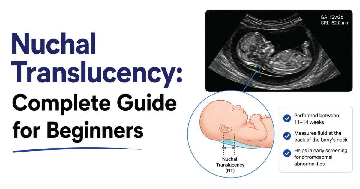
Nuchal translucency refers to the fluid-filled space located at the back of a developing fetus’s neck during the first trimester of pregnancy.
During an ultrasound examination, this fluid appears as a dark or translucent area beneath the skin of the fetal neck.
The thickness of this fluid collection is measured carefully because an increased nuchal translucency measurement may be associated with:
- Chromosomal abnormalities
- Genetic syndromes
- Congenital heart defects
- Certain structural abnormalities
A normal measurement generally indicates a lower risk, while an increased measurement may warrant additional evaluation.
What Is a Nuchal Translucency Scan?
A nuchal translucency scan is a specialized ultrasound examination performed during early pregnancy.
The purpose of the scan is to:
- Measure the thickness of the fetal neck fluid
- Assess fetal development
- Estimate the risk of chromosomal disorders
- Support first-trimester prenatal screening
The scan is non-invasive and poses no known risk to the mother or fetus when performed by qualified healthcare professionals.
When Is the Nuchal Translucency Scan Performed?
The NT scan is typically performed between:
11 weeks and 13 weeks + 6 days of pregnancy
The fetus must have a crown-rump length (CRL) between approximately:
45 mm and 84 mm
This time period is important because the fluid behind the fetal neck can be measured accurately only during this stage of development.
After the first trimester, the fluid is usually absorbed, making the measurement less useful.
Why Is Nuchal Translucency Important?
The nuchal translucency measurement serves as an important prenatal screening tool.
It helps identify pregnancies with an increased risk of:
Chromosomal Abnormalities
Including:
- Down syndrome (Trisomy 21)
- Trisomy 18
- Trisomy 13
- Turner syndrome
Congenital Heart Defects
Some fetuses with increased NT measurements may have heart abnormalities.
Genetic Syndromes
Certain inherited and non-inherited syndromes may be associated with increased nuchal translucency.
Structural Abnormalities
The scan may also suggest the need for further evaluation of fetal anatomy.
Because of these associations, NT screening plays a valuable role in early pregnancy assessment.
How Nuchal Translucency Is Measured
Accurate measurement requires specialized training and strict ultrasound techniques.
Step 1: Positioning the Fetus
The fetus must be viewed in a proper mid-sagittal plane.
Step 2: Magnification
The image is enlarged to allow precise measurement.
Step 3: Identifying the Fluid Space
The sonographer identifies the translucent area behind the fetal neck.
Step 4: Measuring Thickness
The maximum thickness of the fluid is measured using electronic calipers.
Multiple measurements may be obtained to ensure accuracy.
Even small differences in measurement can significantly affect risk calculations.
Normal Nuchal Translucency Measurement
The normal range varies according to gestational age and fetal size.
Generally:
- Measurements below 3.0 mm are often considered within normal limits.
- Most fetuses have measurements between 1 mm and 2.5 mm.
As the fetus grows, the expected NT measurement also increases slightly.
Healthcare providers evaluate results using gestational-age-specific reference charts.
A normal NT measurement usually indicates a lower risk of chromosomal abnormalities.
What Does Increased Nuchal Translucency Mean?
An increased nuchal translucency measurement does not necessarily mean that a fetus has a genetic disorder.
However, it may indicate an increased risk.
Possible associations include:
Down Syndrome (Trisomy 21)
One of the most common conditions associated with increased NT.
Trisomy 18
A serious chromosomal abnormality affecting multiple organ systems.
Trisomy 13
Another chromosomal condition linked to severe developmental abnormalities.
Turner Syndrome
A chromosomal disorder affecting females.
Congenital Heart Disease
Some fetuses with increased NT measurements have normal chromosomes but heart defects.
Genetic Syndromes
Various rare syndromes may also be associated with increased NT.
Further testing is often recommended when the NT measurement is significantly elevated.
Nuchal Translucency and Down Syndrome Screening
The NT scan is widely used as part of first-trimester screening for Down syndrome.
Risk assessment often combines:
- Maternal age
- Nuchal translucency measurement
- Blood test results
- Gestational age
This combined screening approach improves detection rates compared to maternal age alone.
It allows healthcare providers to identify pregnancies that may benefit from additional diagnostic testing.
Combined First-Trimester Screening
Many healthcare providers combine NT measurements with maternal blood tests.
Blood markers commonly evaluated include:
Pregnancy-Associated Plasma Protein A (PAPP-A)
Lower levels may be associated with chromosomal abnormalities.
Free Beta-hCG
Abnormal levels may increase the likelihood of certain conditions.
Combining ultrasound findings with blood tests provides a more accurate risk assessment.
Nuchal Translucency vs Diagnostic Testing
It is important to understand that NT screening is not a diagnostic test.
Screening Test
The NT scan estimates risk.
It does not confirm or exclude a condition.
Diagnostic Test
Diagnostic procedures can provide definitive answers.
Examples include:
- Chorionic Villus Sampling (CVS)
- Amniocentesis
Patients with abnormal screening results may choose diagnostic testing for confirmation.
What Happens If NT Is Increased?
If an increased measurement is identified, healthcare providers may recommend additional evaluation.
Possible next steps include:
Detailed Ultrasound Examination
A comprehensive anatomy scan may be performed later in pregnancy.
Non-Invasive Prenatal Testing (NIPT)
A blood test that analyzes fetal DNA.
Chorionic Villus Sampling (CVS)
Provides direct genetic information.
Amniocentesis
Analyzes fetal chromosomes through amniotic fluid sampling.
Fetal Echocardiography
Evaluates the fetal heart for structural abnormalities.
The appropriate follow-up depends on the degree of risk and patient preferences.
How the NT Scan Is Performed
The procedure is similar to a routine pregnancy ultrasound.
Preparation
Patients may be asked to arrive with a partially full bladder.
Ultrasound Examination
The sonographer places ultrasound gel on the abdomen.
Image Acquisition
The fetus is visualized and measured.
Measurement Recording
The NT thickness is measured and documented.
Risk Calculation
Results may be combined with blood test findings.
The entire examination usually takes 20 to 45 minutes.
Benefits of Nuchal Translucency Screening
NT screening offers several advantages.
Early Risk Assessment
Information becomes available during the first trimester.
Non-Invasive
No needles or surgical procedures are required.
Safe
Ultrasound does not use ionizing radiation.
Improved Pregnancy Management
High-risk pregnancies can receive closer monitoring.
Parental Decision-Making
Results help families make informed healthcare choices.
These benefits make NT screening an important component of prenatal care.
Limitations of Nuchal Translucency Screening
Although valuable, NT screening has limitations.
Not Diagnostic
It cannot confirm a chromosomal condition.
False Positives
Some fetuses with increased NT are completely healthy.
False Negatives
Some abnormalities may occur despite normal NT measurements.
Operator Dependency
Accurate measurements require experienced sonographers.
Therefore, results should always be interpreted within the context of comprehensive prenatal care.
Factors Affecting NT Measurement
Several factors influence measurement accuracy.
Fetal Position
Poor positioning can make measurement difficult.
Gestational Age
Measurements must be obtained during the appropriate time window.
Sonographer Experience
Proper technique is essential.
Image Quality
High-quality equipment improves reliability.
Strict quality standards help ensure accurate results.
Future of Nuchal Translucency Screening
Advances in prenatal imaging continue to improve screening accuracy.
Future developments may include:
- Artificial intelligence-assisted measurements
- Improved ultrasound technology
- Enhanced genetic screening methods
- Earlier risk assessment techniques
- Better integration with prenatal testing programs
Despite advances in genetic testing, NT measurement remains an important component of first-trimester fetal evaluation.
Conclusion
Nuchal Translucency (NT) is a specialized first-trimester ultrasound measurement that evaluates the fluid-filled space at the back of the fetal neck. Performed between 11 and 13 weeks plus 6 days of pregnancy, the examination helps estimate the risk of chromosomal abnormalities such as Down syndrome, Trisomy 18, Trisomy 13, and Turner syndrome, as well as certain congenital heart defects.
Although NT screening is not a diagnostic test, it provides valuable early information that can guide further evaluation and prenatal care. When combined with maternal blood tests and other screening methods, nuchal translucency assessment remains one of the most effective tools for early pregnancy risk assessment and fetal health monitoring.
👉 Learn everything about Echogenicity and what echogenic means in ultrasound imaging. This beginner-friendly guide explains how different tissues reflect sound waves, why structures appear bright on scans, and the clinical significance of echogenic findings. Perfect for students and healthcare professionals. Read the full post to gain a clear understanding of echogenic structures in ultrasound and their diagnostic importance.

Zak is a dedicated medical and career writer specializing in sonography, healthcare education, and professional development. Through SonographerSalary.com, he shares in-depth insights on sonographer salaries, education pathways, and career tips to help readers build successful futures in medical imaging. His content combines accuracy with practical, easy-to-understand guidance, empowering students and professionals to make confident, informed career decisions.