
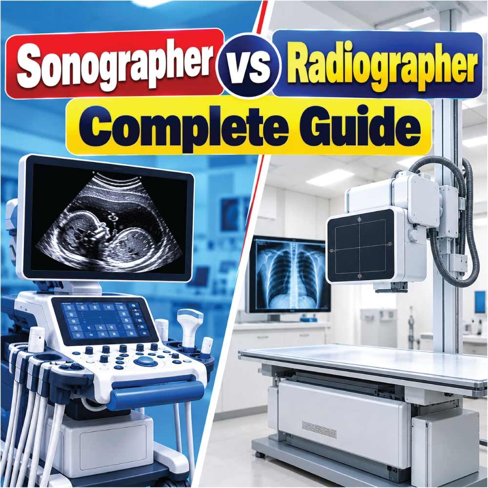
The difference between sonographer and radiographer can be confusing because both professionals work with medical imaging and play an important role in diagnosing and monitoring health conditions. However, they use different technologies, follow different imaging procedures, and often have different day-to-day responsibilities.
A sonographer primarily uses ultrasound technology to create real-time images of structures inside the body. A radiographer, on the other hand, commonly works with X-rays and other forms of medical imaging, depending on their training and role.
Although both careers require technical knowledge, patient-care skills, attention to detail, and an understanding of human anatomy, the working environment can be quite different.
Understanding these distinctions is useful for students considering a healthcare career, patients who want to understand who performs their imaging examination, and anyone researching medical imaging professions.
What Is a Sonographer?
A sonographer is a healthcare professional who uses ultrasound equipment to produce images of internal body structures. The process is known as diagnostic medical sonography or ultrasonography.
During an examination, the sonographer operates an ultrasound machine and uses a handheld device called a transducer. The transducer sends high-frequency sound waves into the body. These waves interact with tissues and return to the equipment, which converts the information into images.
Sonographers may examine many different parts of the body. Their area of specialization can influence the examinations they perform and the patients they work with.
Common sonography specialties include:
- Abdominal sonography
- Obstetric and gynecologic sonography
- Vascular sonography
- Cardiac sonography
- Musculoskeletal sonography
- Breast sonography
- Pediatric sonography
A sonographer does more than simply place a probe on a patient’s body. They select appropriate imaging techniques, adjust equipment settings, obtain diagnostic-quality images, document findings, and communicate relevant information to the appropriate healthcare professionals.
In many healthcare systems, a physician or another qualified provider interprets the completed examination and makes the medical diagnosis.
What Is a Radiographer?
A radiographer is a healthcare professional trained to perform diagnostic imaging examinations, particularly those involving X-rays. The exact scope of practice varies by country, employer, qualification, and specialty.
Radiographers may work with equipment used to produce images of bones, the chest, joints, and other anatomical structures. Depending on their education and professional role, some may also work in areas involving more advanced imaging technologies.
A radiographer’s responsibilities can include preparing patients, positioning them correctly, operating imaging equipment, selecting appropriate technical settings, and ensuring that images meet the required quality standards.
Patient safety is also an important part of radiography. Because X-ray imaging uses ionizing radiation, radiographers are trained in radiation protection principles and techniques designed to minimize unnecessary exposure.
Radiographers may work in:
- Hospitals
- Emergency departments
- Diagnostic imaging centers
- Outpatient clinics
- Specialized medical facilities
- Mobile imaging services
The term used for this profession can also vary internationally. In some regions, professionals performing X-ray examinations may be called radiologic technologists, while “radiographer” is commonly used in other healthcare systems.
Sonographer and Radiographer: Key Differences
The most obvious difference is the imaging technology each professional commonly uses.
| Feature | Sonographer | Radiographer |
|---|---|---|
| Primary technology | Ultrasound | X-ray and related imaging equipment |
| Main energy used | High-frequency sound waves | Ionizing radiation for X-ray imaging |
| Produces real-time imaging | Yes | Generally not in the same way |
| Common examinations | Abdomen, pregnancy, vessels, heart, soft tissues | Chest, bones, joints, and other X-ray examinations |
| Patient positioning | Important | Very important |
| Radiation protection | Not a primary concern for ultrasound | Essential when using ionizing radiation |
| Equipment | Ultrasound machine and transducer | X-ray imaging system and related equipment |
| Common workplace | Hospitals, clinics, imaging centers | Hospitals, imaging centers, clinics |
| Patient interaction | Often substantial | Often substantial |
These are broad distinctions rather than universal rules. The responsibilities of individual professionals can differ according to their specialty, location, credentials, and workplace.
How Their Imaging Technologies Differ
How Ultrasound Works
Ultrasound uses high-frequency sound waves rather than X-rays.
The sonographer applies an ultrasound transducer to the patient’s skin, usually with a conductive gel between the transducer and the skin. The transducer sends sound waves into the body and receives returning echoes.
The ultrasound system processes those echoes to create images.
One major advantage of ultrasound is its ability to provide real-time imaging. This can allow clinicians to observe movement and blood flow and to assess structures dynamically.
Ultrasound is commonly used to examine soft tissues and fluid-filled structures. It is also widely used during pregnancy because it does not use ionizing radiation.
How X-Ray Imaging Works
Radiography uses X-rays, a form of electromagnetic radiation. Different tissues absorb X-rays differently, allowing the imaging system to create a representation of internal structures.
Dense materials such as bone generally absorb more X-rays than surrounding soft tissues, which is why bones can appear prominently on radiographic images.
Radiography is particularly useful for examining:
- Fractures
- Bone alignment
- Chest structures
- Certain joint abnormalities
- Foreign objects
- Other conditions that can be evaluated with projection radiography
The examination technique depends on the body part being imaged and the clinical reason for the examination.
Does a Sonographer Use Radiation?
Diagnostic ultrasound does not use ionizing radiation.
Instead, it uses mechanical sound waves. This is one reason ultrasound is widely used in situations where avoiding ionizing radiation is important.
However, that does not mean every ultrasound examination is automatically appropriate for every situation. The type of imaging selected depends on the patient’s symptoms, clinical question, anatomy, and the healthcare provider’s judgment.
Sonographers must also understand ultrasound safety principles and use appropriate equipment settings and scanning techniques.
Does a Radiographer Use Radiation?
Radiographers who perform X-ray examinations work with ionizing radiation, so radiation protection is a fundamental part of their professional training.
The goal is not simply to produce an image. The examination must be performed appropriately while avoiding unnecessary radiation exposure.
Radiographers may apply principles such as:
- Appropriate patient positioning
- Correct equipment settings
- Proper examination protocols
- Radiation protection procedures
- Careful field selection
- Avoidance of unnecessary repeat images
The exact procedures depend on the imaging system, examination, local regulations, and clinical circumstances.
What Does a Sonographer Do During a Typical Examination?
A sonographer’s work usually involves direct interaction with the patient and the ultrasound system.
A typical examination may involve several stages.
Patient Preparation
The sonographer reviews the examination request and confirms the appropriate patient and procedure. Depending on the examination, the patient may need specific preparation, such as fasting or having a comfortably full bladder.
Preparation requirements vary according to the type of ultrasound.
Patient Positioning
The patient may need to lie down, turn onto a side, sit, or assume another position that provides suitable access to the body area being examined.
Positioning can affect the quality of the examination, so the sonographer must understand anatomy and imaging technique.
Image Acquisition
The sonographer applies gel and moves the transducer over the relevant area.
They adjust settings such as depth, gain, focus, and other controls to obtain useful images. They may also measure anatomical structures or assess blood flow using Doppler ultrasound when appropriate.
Documentation
The sonographer records appropriate images and measurements and documents relevant examination information.
The completed examination is then made available for interpretation according to the healthcare system and professional workflow.
What Does a Radiographer Do During a Typical Examination?
A radiographer also combines technical skills with patient care.
Preparing the Patient
The radiographer confirms the requested examination and explains the procedure. They may ask the patient to remove objects that could interfere with imaging.
Positioning
Correct positioning is particularly important in radiography because an unsuitable position can affect image quality and may require the image to be repeated.
The radiographer must understand anatomical landmarks and the requirements of the specific examination.
Operating the Equipment
The radiographer operates the imaging system and follows the appropriate examination protocol.
Technical factors must be selected carefully to produce an appropriate diagnostic image while following radiation protection principles.
Checking Image Quality
After acquiring an image, the radiographer evaluates whether it meets the necessary technical requirements. If an image does not adequately demonstrate the required anatomy, additional imaging may sometimes be necessary.
Education and Training Differences
The educational pathway for sonographers and radiographers varies significantly between countries.
Both professions generally require formal education in healthcare and medical imaging, but the curriculum reflects their different technologies and responsibilities.
Sonography education commonly emphasizes:
- Human anatomy
- Physiology
- Ultrasound physics
- Patient care
- Imaging techniques
- Pathology
- Doppler principles
- Specialty-specific scanning
Radiography education commonly emphasizes:
- Anatomy and physiology
- Radiographic positioning
- Radiation physics
- Radiation protection
- Patient care
- Image production
- Radiographic procedures
- Pathology
- Equipment operation
Professional registration, certification, licensing, or other regulatory requirements may also apply depending on the jurisdiction.
For anyone considering either career, it is important to check the requirements of the country and state or region where they intend to work.
Sonographer vs Radiographer Work Environment
Both professionals can work in hospitals and outpatient imaging facilities, but their daily environments can differ.
Sonographers often spend considerable time directly beside the patient while performing examinations. Their work may involve prolonged standing and repeated use of the arms and hands while manipulating the transducer.
Radiographers may move between examination rooms and may work with patients who have injuries, mobility limitations, or urgent medical needs. Some radiographers work closely with emergency departments and hospital wards.
The physical demands of both professions can include:
- Standing for extended periods
- Moving or repositioning patients
- Operating specialized equipment
- Maintaining concentration during examinations
- Communicating with patients and healthcare staff
The specific workload depends heavily on the workplace and specialty.
Also Read:
Skills Needed for Each Career
Important Sonographer Skills
Sonography combines technical knowledge with hands-on scanning ability.
Important skills include:
- Strong understanding of anatomy
- Hand-eye coordination
- Spatial awareness
- Attention to detail
- Communication
- Patient positioning
- Ultrasound physics
- Ability to recognize relevant anatomical structures
- Ability to adapt scanning techniques
Sonographers also need patience because some examinations require careful scanning from multiple angles.
Important Radiographer Skills
Radiographers require a strong technical foundation as well.
Useful skills include:
- Knowledge of anatomy
- Accurate positioning
- Understanding of radiation protection
- Equipment operation
- Attention to detail
- Communication
- Problem-solving
- Ability to follow imaging protocols
- Patient-care skills
Radiographers must also be able to work efficiently without allowing speed to compromise patient safety or image quality.
Sonographer and Radiographer Specialties
The range of specialties is another important difference.
Common Sonography Specialties
A sonographer may develop expertise in areas such as:
| Specialty | Typical Focus |
|---|---|
| Abdominal | Liver, gallbladder, kidneys, pancreas and related structures |
| Obstetric | Pregnancy and fetal assessment |
| Gynecologic | Female reproductive organs |
| Vascular | Blood vessels and blood flow |
| Cardiac | Heart structure and function |
| Breast | Breast tissue assessment |
| Musculoskeletal | Muscles, tendons, ligaments and related structures |
| Pediatric | Ultrasound examinations involving children |
The exact scope and terminology can differ between employers and professional organizations.
Common Radiography Areas
Radiography can also lead to different areas of practice, depending on the qualification and healthcare system.
These may include:
- General diagnostic radiography
- Emergency imaging
- Orthopedic imaging
- Mobile radiography
- Operating-room imaging
- Specialized imaging pathways
Some radiographers pursue additional education to work with modalities such as computed tomography or magnetic resonance imaging, where permitted by their professional training and local regulations.
Also Read:
Which Patients Do They Commonly See?
Both professions can work with a very broad patient population.
A sonographer may examine:
- Pregnant patients
- Adults with abdominal symptoms
- Patients undergoing vascular assessments
- Children
- Patients requiring cardiac imaging
- People being evaluated for soft-tissue conditions
Radiographers may image:
- Patients with suspected fractures
- People with chest symptoms
- Emergency patients
- Surgical patients
- Orthopedic patients
- Patients requiring routine diagnostic imaging
Neither profession is limited to one type of patient.
Sonographer vs Radiographer: Patient Interaction
Patient communication is important in both careers.
A sonographer may spend a relatively long period with a patient during a detailed examination. They may need to explain positioning, breathing instructions, and what the patient should expect during scanning.
Radiographers also communicate directly with patients, but individual interactions can vary considerably depending on the examination.
For example, a patient receiving an X-ray may have a relatively short imaging procedure, while a complex ultrasound examination can require more prolonged scanning.
The amount of patient interaction therefore depends not only on the profession but also on the specialty and workplace.
Also Read:
Can a Radiographer Become a Sonographer?
A radiographer may be able to transition into sonography, but this is not automatically available simply because both careers involve medical imaging.
Additional education and clinical training in ultrasound are generally required.
The exact pathway depends on:
- Country or region
- Existing qualification
- Professional registration
- Employer requirements
- Sonography program availability
- Specialty chosen
A radiographer considering this transition should check the current educational and registration requirements in the jurisdiction where they intend to practice.
Can a Sonographer Become a Radiographer?
A sonographer may also be able to move into radiography, but the two professions have different technical foundations.
Someone trained primarily in ultrasound would normally need additional education in radiographic imaging, radiation physics, positioning, radiation protection, and clinical practice.
Existing healthcare education may provide useful background, but it does not necessarily replace the training required for professional radiography practice.
Career Considerations for Students
Students choosing between sonography and radiography should look beyond job titles and consider what type of imaging work they actually enjoy.
Sonography may appeal to someone interested in:
- Real-time imaging
- Hands-on scanning
- Anatomy and physiology
- Direct patient interaction
- Ultrasound physics
- Developing expertise in a specific sonography specialty
Radiography may appeal to someone interested in:
- X-ray imaging
- Medical imaging technology
- Patient positioning
- Radiation protection
- Working across different clinical environments
- Building a broader imaging career pathway
Neither description applies equally to every workplace, so researching local training programs and speaking with practicing professionals can provide useful insight.
Sonographer and Radiographer Salary Considerations
Salary should be evaluated using current information for the specific country, region, specialty, experience level, and employer.
There is no single worldwide salary figure that accurately represents either profession.
Pay can be influenced by factors such as:
- Professional qualifications
- Years of experience
- Specialty
- Hospital versus outpatient employment
- Geographic location
- Shift patterns
- Additional certifications
- Seniority
- Public or private healthcare employment
Students should therefore compare local salary data rather than relying on generalized international figures.
Sonographer vs Radiographer: Quick Comparison
The following summary brings the main distinctions together.
| Category | Sonographer | Radiographer |
|---|---|---|
| Main imaging method | Ultrasound | X-ray-based radiography |
| Ionizing radiation | No | Yes, when performing X-ray imaging |
| Main device | Transducer and ultrasound system | X-ray imaging equipment |
| Real-time imaging | Major feature | Not the typical defining feature |
| Soft-tissue imaging | Common | More limited with conventional X-ray |
| Bone imaging | Limited compared with X-ray | Common |
| Radiation safety training | Not centered on ionizing radiation | Core component of X-ray practice |
| Hands-on scanning | Extensive | Positioning-focused |
| Patient interaction | Often substantial | Often substantial |
| Specialty options | Abdominal, vascular, cardiac, obstetric and others | General radiography and further imaging pathways |
| Work settings | Hospitals, clinics, imaging centers | Hospitals, clinics, imaging centers |
The table provides a broad comparison. Actual responsibilities can vary depending on professional scope and local healthcare regulations.
Why Both Professions Matter in Modern Healthcare
Medical imaging is not one single discipline. Different technologies answer different clinical questions.
Ultrasound can provide dynamic information about tissues, fluid, movement, and blood flow. Radiography can provide valuable images of bones, the chest, and other structures that respond well to X-ray imaging.
In many healthcare settings, these professionals work as part of a larger clinical team that may include physicians, nurses, radiologists, surgeons, and other specialists.
The imaging professional’s role is important because high-quality diagnostic information depends not only on the equipment but also on how effectively the examination is performed.
Final Thoughts on the Difference Between Sonographer and Radiographer
The central difference between sonographer and radiographer is the imaging technology and professional training involved.
A sonographer primarily uses ultrasound and sound waves to create real-time images, while a radiographer commonly performs X-ray examinations using ionizing radiation. Their education, equipment, safety considerations, examination techniques, and specialty options reflect these differences.
Both careers combine technology with patient care. Neither professional simply operates a machine. They must understand anatomy, communicate with patients, follow appropriate procedures, and produce useful diagnostic images.
For students choosing a career, the most useful comparison is therefore not simply the job title. Consider the type of technology, patient interaction, physical work, education pathway, specialty opportunities, and professional requirements that fit your interests.
Checking the requirements of the relevant professional body or licensing authority is also essential because training and scope of practice can vary by location.

Zak is a dedicated medical and career writer specializing in sonography, healthcare education, and professional development. Through SonographerSalary.com, he shares in-depth insights on sonographer salaries, education pathways, and career tips to help readers build successful futures in medical imaging. His content combines accuracy with practical, easy-to-understand guidance, empowering students and professionals to make confident, informed career decisions.