
In ultrasound imaging, the terms hypoechoic and hyperechoic are crucial for interpreting tissue characteristics. They describe how structures reflect sound waves, influencing their brightness on grayscale ultrasound images. Understanding these concepts is essential for diagnosing medical conditions, identifying lesions, and guiding procedures accurately.
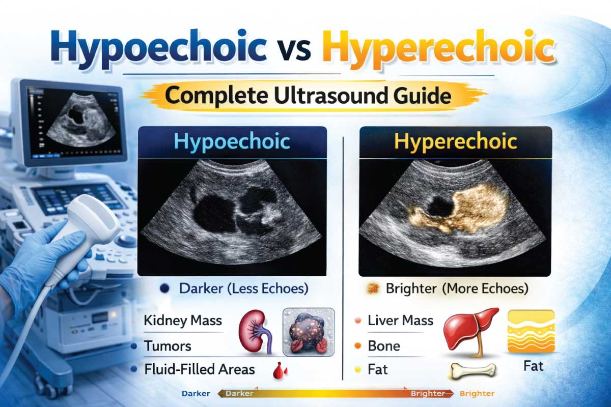
Hypoechoic and hyperechoic structures provide different information about tissues. Hypoechoic areas appear darker because they reflect fewer sound waves, while hyperechoic areas appear brighter due to stronger echoes. Clinicians rely on these patterns to differentiate normal tissues from pathological changes and assess disease severity.
Understanding Hypoechoic Ultrasound
Hypoechoic refers to tissues or structures that reflect fewer ultrasound waves, appearing darker than surrounding tissues on a grayscale image. These structures absorb or transmit most of the sound waves rather than reflecting them back.
Common hypoechoic areas include muscles, certain solid masses, lymph nodes, or fluid-poor tumors. Identifying hypoechoic regions helps clinicians detect inflammation, tumors, cysts, or edema, which may require further evaluation using complementary imaging modalities or laboratory tests.
Understanding Hyperechoic Ultrasound
Hyperechoic refers to tissues that reflect more sound waves, appearing brighter on ultrasound images. These areas produce strong echoes due to high density, interface boundaries, or calcified tissue components.
Examples include bone, fat, fibrous tissue, gallstones, and calcifications. Hyperechoic regions are valuable in detecting tissue fibrosis, vascular plaques, or tumors with calcification, guiding accurate diagnosis and therapeutic interventions.
How Hypoechoic and Hyperechoic Patterns Form
Ultrasound waves travel through tissues at different speeds. When they encounter a difference in acoustic impedance between tissues, some sound is reflected, creating echoes.
- Hypoechoic structures → weak reflection, absorb more sound → darker appearance
- Hyperechoic structures → strong reflection, reflect most sound → brighter appearance
The contrast between hypoechoic and hyperechoic areas allows clinicians to distinguish normal tissue, cysts, solid masses, or calcified structures.
Factors Affecting Echogenicity
| Factor | Effect on Echogenicity | Example |
|---|---|---|
| Tissue composition | Denser tissues are hyperechoic | Bone, fibrous tissue |
| Fluid content | High fluid → hypoechoic | Cysts, abscesses |
| Frequency of ultrasound | Higher frequency → better resolution | Superficial organs |
| Angle of incidence | Perpendicular waves → stronger echoes | Vascular walls |
| Pathological changes | Fibrosis → hyperechoic, edema → hypoechoic | Liver cirrhosis, inflammation |
These factors influence the interpretation of hypoechoic and hyperechoic areas in clinical imaging.
Clinical Applications of Hypoechoic Structures
Hypoechoic regions are common in various diagnostic applications:
1. Abdominal Imaging
- Liver lesions (hypoechoic masses) may indicate tumors or cysts
- Kidney hypoechoic areas suggest solid lesions or inflammation
- Pancreatic hypoechoic regions can indicate pancreatitis or tumors
Identifying hypoechoic structures helps determine whether further testing or intervention is required.
2. Obstetrics and Gynecology
- Ovarian cysts often appear hypoechoic
- Certain uterine masses may be hypoechoic compared to surrounding tissue
- Fetal organs like the liver or kidneys may show hypoechoic patterns in development
These patterns guide diagnosis, monitoring, and treatment decisions in gynecology and obstetrics.
3. Musculoskeletal Ultrasound
- Muscle tissue generally appears hypoechoic relative to tendons
- Fluid collections such as hematomas or bursitis are also hypoechoic
- Hypoechoic masses may indicate tumors or inflammation
Sonographers use hypoechoic patterns to detect tears, inflammation, or tumors.
Clinical Applications of Hyperechoic Structures
Hyperechoic areas also play a key role in diagnosis:
1. Abdominal Imaging
- Gallstones are highly hyperechoic with posterior shadowing
- Fatty infiltration of organs may increase echogenicity
- Liver fibrosis and cirrhosis show diffuse hyperechoic patterns
These findings are crucial for diagnosing metabolic or structural diseases.
2. Cardiac Imaging
- Heart valves are hyperechoic due to dense fibrous tissue
- Calcifications in the myocardium or vessels appear bright
- Echocardiography uses these patterns to assess valve function and detect pathology
3. Musculoskeletal Imaging
- Tendons and ligaments appear hyperechoic relative to muscles
- Bone surfaces and calcifications are extremely hyperechoic
- Identifying hyperechoic lesions helps detect bone spurs, calcific tendinitis, or scar tissue
Comparison of Hypoechoic and Hyperechoic Structures
| Feature | Hypoechoic | Hyperechoic |
|---|---|---|
| Appearance | Dark gray or black | Bright white |
| Reflection | Weak | Strong |
| Common examples | Muscle, cysts, tumors | Bone, fat, calcifications |
| Clinical relevance | Indicates soft tissue lesions or fluid | Indicates dense tissue, fibrosis, or calcifications |
| Diagnostic value | Useful for detecting masses, inflammation | Useful for detecting plaques, stones, or scarring |
Both hypoechoic and hyperechoic structures are essential for differentiating pathology from normal tissue.
Discover More:
Echogenicity Patterns in Lesions
Understanding echogenicity helps classify lesions:
- Hypoechoic mass → solid lesion, inflammation, or tumor
- Hyperechoic mass → fibrous tissue, calcifications, or fat-containing lesion
- Mixed echogenicity → heterogeneous tumors, complex cysts
Accurate interpretation guides biopsy, treatment planning, and monitoring.
Role of Hypoechoic and Hyperechoic Patterns in Ultrasound Diagnosis
- Hypoechoic regions often suggest soft tissue pathology
- Hyperechoic regions indicate dense tissue, calcifications, or fibrosis
- Comparing echogenicity with surrounding tissues enhances diagnostic accuracy
- Patterns assist in identifying cysts, solid tumors, abscesses, or vascular plaques
This comparative approach is essential for precision in ultrasound imaging.
Advanced Applications
1- Quantitative Echogenicity
Modern ultrasound software can measure pixel intensity to quantify hypoechoic and hyperechoic areas. This is useful for:
- Liver fibrosis assessment
- Tumor characterization
- Monitoring disease progression
2- Doppler Ultrasound Integration
Doppler imaging combined with echogenicity helps evaluate:
- Blood flow within hypoechoic or hyperechoic masses
- Vessel obstruction or stenosis
- Tumor vascularity
These applications increase diagnostic precision and treatment planning.
Discover More:
Advantages of Understanding Hypoechoic vs Hyperechoic
| Advantage | Explanation |
|---|---|
| Accurate lesion identification | Differentiates cystic vs solid masses |
| Non-invasive | Provides real-time imaging without radiation |
| Guiding procedures | Assists in biopsy or drainage |
| Disease monitoring | Tracks progression of liver, cardiac, or musculoskeletal conditions |
| Enhances diagnostic confidence | Enables precise assessment of abnormal tissue |
Recognizing echogenicity patterns is essential for safe and effective patient care.
Limitations of Hypoechoic and Hyperechoic Assessment
| Limitation | Reason |
|---|---|
| Operator-dependent | Requires experience to interpret accurately |
| Subjective | Visual brightness assessment may vary |
| Limited specificity | Similar echogenicity can represent different pathologies |
| Obscured visualization | Gas, bone, or deep structures may hinder imaging |
Despite these limitations, echogenicity remains a core principle of ultrasound interpretation.
Tips for Accurate Assessment
- Use appropriate transducer frequency for target depth
- Adjust gain settings to enhance contrast
- Compare the lesion to adjacent normal tissue
- Use multiple planes for a complete view
- Combine with Doppler or elastography if needed
Following these tips ensures accurate hypoechoic and hyperechoic differentiation.
Echogenicity Patterns by Organ
Liver
- Hypoechoic → lesions or edema
- Hyperechoic → fatty infiltration or fibrosis
Kidney
- Cortex is hypoechoic relative to liver
- Hyperechoic medulla may indicate calcifications
Thyroid
- Nodules may appear hypoechoic or hyperechoic
- Homogeneous thyroid tissue is isoechoic
Breast
- Solid tumors are often hypoechoic
- Fatty tissue and fibrous bands are hyperechoic
Heart
- Valves and dense fibrous structures are hyperechoic
- Blood-filled chambers are hypoechoic
Discover More:
Conclusion
Understanding hypoechoic vs hyperechoic patterns is essential for accurate ultrasound interpretation. Hypoechoic areas indicate soft tissue, fluid, or lesions, while hyperechoic areas indicate dense tissue, calcifications, or fibrosis.
Correct interpretation improves diagnostic accuracy, guides procedures, and helps monitor disease progression. By mastering these echogenicity concepts, sonographers and clinicians can deliver precise, safe, and effective patient care.
Hypoechoic and hyperechoic assessments, combined with Doppler and other advanced ultrasound techniques, remain cornerstones of modern diagnostic imaging, ensuring that clinicians detect abnormalities and plan treatments confidently.

Zak is a dedicated medical and career writer specializing in sonography, healthcare education, and professional development. Through SonographerSalary.com, he shares in-depth insights on sonographer salaries, education pathways, and career tips to help readers build successful futures in medical imaging. His content combines accuracy with practical, easy-to-understand guidance, empowering students and professionals to make confident, informed career decisions.