
When you hear the term ultrasound, many of us immediately picture a black-and-white image on a screen, perhaps from a prenatal visit or a diagnostic scan. But what about sonography? Are they the same thing or different? Often these terms are used interchangeably, but there are subtle distinctions that matter, especially if you’re a patient, a budding sonography student, or simply curious about medical imaging. In this comprehensive guide, we will walk you through everything you need to know about sonography and ultrasound, what they are, how they overlap, and why the difference matters. By the end, you will feel confident using the right term, understanding the technology, and knowing when and why these scans are performed.
What Is Ultrasound?
Ultrasound refers to high-frequency sound waves, above the range of human hearing, that travel through the body and reflect off tissues, organs, or fluids. A machine then captures these echoes and converts them into images or videos that radiologists, sonographers, or doctors can interpret. Because ultrasound waves are safe (no ionizing radiation) and noninvasive, ultrasound imaging has become a go-to diagnostic tool across many areas: obstetrics, cardiology, orthopedics, abdominal imaging, and more. In short, ultrasound is the core imaging technology — the physics — behind many scans.
What Is Sonography?
Sonography, on the other hand, refers to the practice or process of using ultrasound to produce images, plus the broader field of work involving interpretation, operation of the equipment, and interaction with patients. A professional who performs sonography — a sonographer — must know how to position the transducer, adjust settings for the best resolution, interpret the images, and often liaise with doctors about what the images show. In many ways, sonography is the application of ultrasound.
So while ultrasound is the technology, sonography is the craft, expertise, and process that makes use of it.
The Relationship Between Ultrasound and Sonography
Given the definitions above, it’s easy to see why the terms are often used interchangeably, but it also helps clarify when precision matters. If you say “ultrasound,” you might be referring to the technique, the waves themselves, or the resulting image. If you say “sonography,” you are more likely referring to the process — including the skill of the person performing the scan. In practice:
A patient receives an “ultrasound scan.”
The person performing it is a “sonographer.”
The field or profession is “sonography.”
Understanding the distinction is important when considering education and careers, discussing limitations and accuracy, or even reviewing a medical report.
📌 What Is Sonography? The Expert Complete Guide explains the science, applications, and career opportunities in medical imaging. This post breaks down techniques, benefits, and real-world uses of sonography. Read the full guide to gain a deep understanding and discover how to start or advance your career in this growing field.
Equipment and Technology: How Ultrasound Machines Work
To appreciate sonography, it helps to understand the equipment. At a basic level, an ultrasound machine includes a transducer (probe), a processing unit, and a display. The transducer sends out high-frequency sound waves and receives echoes when those waves bounce back. The processing unit converts those echoes into grayscale or color-coded images, while the display presents them in real time.
The frequency of sound waves matters: higher frequencies (e.g., 7–15 MHz) yield higher resolution images but penetrate only shallow depths — ideal for superficial structures like tendons or breast tissue. Lower frequencies (e.g., 2–5 MHz) penetrate deeper (e.g., abdomen or pelvis) but at lower resolution.
| Frequency (MHz) | Common Use | Typical Penetration Depth |
|---|---|---|
| 7–15 | Superficial structures (thyroid, breast, musculoskeletal) | ~2–5 cm |
| 5–7 | Medium-depth imaging (small organs, veins) | ~4–8 cm |
| 2–5 | Deep organs (liver, kidneys, fetal imaging) | ~8–15 cm |
Different ultrasound machines also offer specialized modes:
| Mode | Purpose |
|---|---|
| 2D (B-mode) | Standard grayscale “slice” images of anatomy |
| Doppler | Assesses blood flow velocity and direction (vascular studies) |
| Color-flow Doppler | Visualizes blood flow in color overlaid on 2D images |
| 3D/4D Ultrasound | Three-dimensional imaging, often used for fetal anatomy or volume measurement |
| Elastography | Estimates tissue stiffness (useful in liver, thyroid, breast imaging) |
This variety allows sonography to be tailored to numerous diagnostic needs, making ultrasound technology remarkably versatile.
Types of Sonography / Ultrasound Exams
Sonography isn’t a one-size-fits-all procedure. Depending on what part of the body needs imaging and what question needs answering, different types of ultrasound exams are used. For example:
Abdominal ultrasound — for liver, gallbladder, kidneys, pancreas
Pelvic ultrasound — for uterus, ovaries, prostate, bladder
Obstetric ultrasound — for fetal monitoring during pregnancy
Vascular ultrasound — for blood flow in arteries and veins
Cardiac ultrasound (echocardiography) — for heart structure and function
Musculoskeletal ultrasound — for muscles, tendons, joints
Thyroid or neck ultrasound — for glands and lymph nodes
| Exam Type | Typical Indications | Special Considerations |
|---|---|---|
| Abdominal | Pain, swelling, abnormal lab tests | Patient may need to fast for better clarity |
| Pelvic | Pelvic pain, menstrual disorders, infertility | Full bladder sometimes required |
| Obstetric | Prenatal screening, fetal growth, anatomy checks | Timing with gestational age matters |
| Vascular (Doppler) | DVT, varicose veins, arterial narrowing, clots | Patient must remain still; sometimes compression |
| Cardiac (Echocardiogram) | Heart murmurs, CHF, valve issues, congenital defects | Requires skilled sonographer; multiple views |
| Musculoskeletal | Sprains, tears, inflammation, fluid accumulation | High-frequency probe; often dynamic imaging |
| Thyroid/Neck | Nodules, goiter, lymph node enlargement | Patient’s neck position matters |
As you can see, sonography touches nearly every specialty — making it a cornerstone of modern diagnostic imaging.
📌 Diagnostic Imaging Courses Online offer a flexible way to gain essential skills in medical imaging from anywhere. This post explores course options, benefits, and career opportunities. Read the full article to find the best programs and start building a successful career in diagnostic imaging today.
Who Performs Sonography? The Role of a Sonographer
A key difference between ultrasound and sonography lies in the human element. The person performing the exam — the sonographer — plays a critical role. A sonographer must not only know how to operate the machine, but also how to position the transducer, optimize settings (gain, depth, focus), and sometimes even guide a patient into a better posture to improve image quality.
Sonographers also act as patient liaisons. They explain preparation instructions, reassure anxious patients (especially in obstetrics), and sometimes provide immediate feedback to doctors when urgent findings arise. Their skill level greatly affects the diagnostic accuracy — a poorly performed scan can lead to missed abnormalities or misleading results.
For those interested in a medical imaging career, entering the field of sonography typically involves formal training (certificate, associate, or bachelor’s program), clinical hours, and possibly certification depending on the country.
Sonography vs Ultrasound: Common Confusions and Clarifications
Because the terms overlap in casual usage, many people confuse them. A few common misconceptions:
Thinking that “ultrasound” always refers to pregnancy scans. In reality, pregnancy is just one of many applications.
Assuming “sonography” and “ultrasound” are interchangeable with “radiography” or “X-ray imaging.” That’s incorrect — ultrasound doesn’t use ionizing radiation.
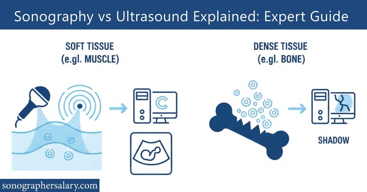
Believing ultrasound is always superior for all diagnostic imaging — yet sometimes other modalities (CT, MRI) are better for certain conditions like bone fractures or detailed tissue contrast.
Understanding these nuances helps patients ask informed questions, helps medical students choose correct terminology, and helps maintain clarity in medical documentation.
Advantages and Limitations of Sonography / Ultrasound
Like any medical tool, ultrasound and sonography come with strengths and weaknesses. Recognizing them helps set realistic expectations.
| Advantages | Limitations |
|---|---|
| Safe (no ionizing radiation) | Limited penetration for deep or air-filled structures |
| Real-time imaging (movement, blood flow) | Image quality dependent on operator skill and patient body habitus |
| Relatively low cost compared to CT/MRI | Cannot image bones or gas-filled organs well |
| Portable — can be used bedside or in remote clinics | Limited tissue contrast compared to MRI |
| Wide variety of uses (abdomen, heart, OB, vascular, MSK) | Sometimes less definitive — may require follow-up with other imaging |
For example, ultrasound is excellent for viewing a fetus, gallbladder stones, or detecting fluid in a joint but if you suspect a complex bone fracture, a CT scan or X-ray may be far more revealing.
📌 High-Paying Medical Jobs Without a Degree reveal rewarding healthcare careers you can start quickly. This post highlights top roles, salaries, and how to enter the field without a formal degree. Read the full guide to discover practical steps to launch a lucrative medical career today.
Practical Tips for Patients Preparing for a Sonography Exam
If you’re scheduled for a sonogram, here are a few friendly practical tips to ensure smooth results. Often, small details on your end make a big difference in image clarity.
First, follow preparation instructions carefully. Depending on the exam type, you might be asked to drink water (to fill your bladder), fast, or wear loose clothing. Drink enough water but don’t overhydrate — sometimes a slightly overfull bladder creates discomfort or poor images.
Second, arrive slightly early and ask questions. If you don’t understand what you need to do (e.g., holding breath, changing posture), don’t hesitate to ask the sonographer.
Third, relax. Tensing up muscles or being anxious can interfere with image acquisition — especially in musculoskeletal or vascular scans.
In simple terms: prepare properly, communicate clearly, and stay calm — that’s the recipe for optimal sonography results.
Common Mistakes People Make Regarding Sonography / Ultrasound
Even though ultrasound is widely used and seemingly straightforward, mistakes happen. A few common errors or misunderstandings include:
Going in underdressed or wearing tight garments when loose clothing was advised.
Eating before a scan when fasting was required, which can muddy abdominal imaging.
Forgetting to drink water when a full bladder was needed for clear pelvic visualization.
Assuming a normal scan rules out all disease — ultrasound has limitations, and some conditions may require follow-up with MRI, CT, or biopsy.
Using low-frequency probes for superficial structures, resulting in poor resolution.
Avoiding these mistakes helps guarantee the best possible outcome from your scan.
Interpreting Results and When Follow-up Is Needed
After your exam, the sonographer’s images and measurements are usually reviewed by a radiologist or relevant specialist. Their report may describe what was visualized (e.g., gallstones, fetal biometric measurements, or blood flow speed) and note whether anything is abnormal.
It’s helpful to remember that a normal ultrasound result does not always guarantee absence of disease. For instance, small lesions or early-stage conditions may not be visible due to ultrasound’s resolution limits or because they lie behind bone or gas. If your symptoms persist or labs remain abnormal despite a “clean” scan, doctors may recommend further imaging — such as MRI, CT, or functional studies — or repeat ultrasound later.
Also, keep in mind that normal “ranges” depend on many factors: the machine’s resolution, the operator’s skill, patient’s body type, and the organ or tissue being scanned. Don’t hesitate to discuss with your doctor whether ultrasound is sufficient or if more sensitive imaging is needed.
Cost, Accessibility, and Global Reach of Sonography
One of the biggest benefits of ultrasound is its relative affordability compared to high-end imaging modalities like MRI or CT. Because equipment tends to be less expensive, portable, and energy-efficient, ultrasound machines are common not only in major hospitals but also in small clinics, rural health centers, and mobile health units. This makes sonography a powerful tool for early detection, maternal care, and diagnostics in remote or resource-limited settings.
| Setting | Typical Cost Range (USD equivalent) | Common Use Cases |
|---|---|---|
| Urban hospital (full facility) | $50–$200 per scan | Obstetrics, abdominal, cardiac, vascular |
| Small clinic / outpatient lab | $20–$80 per scan | Routine checkups, prenatal screening |
| Mobile health unit / rural clinic | $5–$40 per scan | Maternal care, basic diagnostics |
| Emergency / bedside ultrasound | Included in hospital service | Trauma, urgent fluid detection |
Because of this accessibility and low cost, sonography plays a vital role globally — from urban centers to remote villages. Its portability also means it can be used in emergency rooms, ambulances, or remote camps, making it invaluable in crisis or low-resource environments.
Future Trends in Sonography Technology
Sonography continues to evolve at a rapid pace. Emerging advancements promise to make ultrasound even more powerful and widely accessible:
Portable handheld scanners: Compact, smartphone-connected devices are making point-of-care ultrasound more common, even in field clinics or at-home visits.
AI-assisted image analysis: Artificial intelligence tools increasingly help detect abnormalities — for example, automated measurements of fetal size, detection of tumors, or blood flow irregularities.
Improved resolution and penetration: New probe designs and signal processing are pushing the boundaries of what can be visualized, possibly challenging imaging modalities like MRI in certain uses.
Integration with telemedicine: Remote sonography consultations, where scans are performed locally and interpreted by specialists elsewhere, are gaining traction — especially in underserved areas.
These innovations are set to expand the scope of sonography, making it faster, more accurate, and more accessible than ever before.
📌 Sonographer Salary in USA breaks down earnings, factors affecting pay, and career growth opportunities for sonographers nationwide. This post provides real insights and practical tips for maximizing your income. Read the full guide to understand what you can earn and how to advance in this in-demand healthcare profession.
When Sonography Is not Enough: Recognizing the Limits
It’s tempting to assume that because ultrasound is versatile and widely available, it should be the first (or only) diagnostic tool. However, not every condition can or should be diagnosed with ultrasound.
For instance, bone disorders, air-filled organs (like the lungs), or tiny lesions deep within tissues often require other imaging methods. CT or MRI provide higher contrast for soft-tissue differentiation, bone detail, or intricate anatomical mapping. Sometimes, a biopsy or functional test may be necessary.
Also, ultrasound image quality can degrade in patients with certain body types (e.g., very obese patients) or due to interferences like gas or scarring. In those cases, alternate imaging might give a clearer answer.
Using sonography as part of a broader diagnostic strategy rather than sole reliance often delivers better patient care.
Why Terminology Matters: Professionalism, Clarity, and Career
You might wonder — does it really matter whether you say “ultrasound” or “sonography”? The answer: yes, especially in a professional or educational context.
For healthcare professionals, using precise language supports clear documentation and communication. A medical student learning the trade benefits from knowing that sonography refers to the practice and skill of imaging, not just the equipment or the scan. For patients, understanding the terms helps ask better questions: about who performs the scan, their qualification, the equipment used, and whether follow‑up imaging is likely needed.
For those considering a career in medical imaging, the distinction clarifies what training is required, what job roles exist, and what skills you’ll need to succeed.
Final Thoughts: Sonography and Ultrasound — Two Sides of the Same Coin
At its core, ultrasound is a safe, versatile, and powerful imaging technology using high‑frequency sound waves. Sonography is the human-driven art and science of leveraging that technology to help diagnose, monitor, and sometimes guide treatment. Together they form one of the most widely used, cost-effective, and accessible diagnostic tools in modern medicine.
For patients, being informed about what sonography can and cannot, do helps set realistic expectations. For aspiring medical professionals, understanding the terminology and technology lays a foundation for meaningful training and responsible practice.
Whether you are scheduling your first ultrasound, studying to become a sonographer, or simply curious about how medical imaging works, you are now better equipped to understand the language, science, and human skill behind the images.
Conclusion
Ultrasound and sonography share a close bond: one is the technology, the other is the skilled practice. While ultrasound provides the waves and images, sonography brings human expertise, precision, and care. Throughout this guide we have explored how the two relate, how they are applied across different medical fields, what advantages they offer, and where they fall short. We have also offered practical tips for patients and shed light on emerging trends in the field.
Understanding this distinction not only empowers you as a patient or informed reader, but also supports better communication in healthcare, clearer documentation, and responsible use of diagnostic tools. If you’re considering a sonography exam or a career in medical imaging keep this guide handy. And remember: the next time someone says “ultrasound scan,” you will know exactly what’s behind that simple phrase.
FAQs:
Are sonography and ultrasound the same?
Sonography and ultrasound are closely related but not identical. Ultrasound refers to the technology using high-frequency sound waves, while sonography is the practice of performing scans with that technology. Essentially, ultrasound is the tool, and sonography is the process of producing images for diagnosis.
What are the three types of ultrasound?
The three main types of ultrasound are: 2D ultrasound, which produces flat images; 3D ultrasound, giving detailed three-dimensional images; and Doppler ultrasound, which measures blood flow. Each type has specific uses, from routine pregnancy checks to diagnosing heart or vascular conditions accurately.
Is sonography used to detect pregnancy?
Yes, sonography is commonly used to detect pregnancy. It confirms gestational age, monitors fetal growth, checks heartbeat, and identifies potential complications. Early pregnancy scans help ensure healthy development, while later scans guide medical decisions and provide expectant parents with visual updates on their baby’s progress.
Can a sonographer do ultrasound?
Yes, a sonographer is trained to perform ultrasound scans. They operate the equipment, capture images, and sometimes provide preliminary interpretations for doctors. Sonographers specialize in areas like obstetric, cardiac, or vascular imaging, ensuring safe, accurate, and detailed diagnostic results for patient care.
What exactly is sonography?
Sonography is a medical imaging technique that uses high-frequency sound waves to create real-time images of the body’s internal structures. It is non-invasive, safe, and widely used for monitoring pregnancy, examining organs, evaluating blood flow, and guiding procedures, providing essential information for accurate medical diagnosis and treatment.
What is another name for ultrasound?
Another name for ultrasound is sonography or ultrasonography. All terms refer to the use of high-frequency sound waves to create images of internal body parts. These techniques are widely applied in medicine for pregnancy monitoring, organ evaluation, and diagnosing conditions without the use of harmful radiation.

Zak is a dedicated medical and career writer specializing in sonography, healthcare education, and professional development. Through SonographerSalary.com, he shares in-depth insights on sonographer salaries, education pathways, and career tips to help readers build successful futures in medical imaging. His content combines accuracy with practical, easy-to-understand guidance, empowering students and professionals to make confident, informed career decisions.