
Ultrasonography, often called ultrasound imaging, is one of the most widely used diagnostic tools in modern medicine. It allows healthcare professionals to see inside the human body in real time without using radiation, making it safe, effective, and versatile.
From monitoring pregnancy to diagnosing heart and organ conditions, ultrasonography plays a crucial role in patient care.
In this article, we will explore how ultrasonography works, its uses, benefits, and career opportunities in this growing medical field.
What is Ultrasonography?
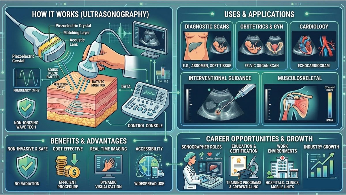
Ultrasonography is a medical imaging technique that uses high-frequency sound waves to create images of structures inside the body. These sound waves are emitted by a device called a transducer, which also receives the reflected waves and converts them into visual images displayed on a screen.
Unlike X-rays or CT scans, ultrasonography does not use ionizing radiation. This makes it safer for patients, especially pregnant women and children. It is commonly used in hospitals, clinics, and diagnostic centers for both routine checkups and emergency diagnostics.
How Does Ultrasonography Work?
The process of ultrasonography is based on the reflection of sound waves. When the transducer is placed on the skin, it sends sound waves into the body. These waves travel through tissues and bounce back when they hit different structures such as organs or fluids.
The returning echoes are captured by the transducer and sent to a computer, which processes them into images. These images help doctors analyze the size, shape, and condition of internal organs.
Key Steps in the Process
- The technician applies a gel to the skin to improve sound wave transmission
- The transducer is moved over the targeted area
- Sound waves penetrate the body and reflect back
- A computer converts echoes into real-time images
This process is painless, quick, and non-invasive, making it highly preferred in medical diagnostics.
Types of Ultrasonography
Ultrasonography includes several specialized types, each designed for specific diagnostic purposes. These techniques help doctors examine different organs and systems in the body with precision. From monitoring pregnancy to assessing heart health, each type of ultrasound provides valuable insights. Understanding these types helps in choosing the right imaging method for accurate diagnosis and effective treatment.
1. Abdominal Ultrasound
Abdominal ultrasound is commonly used to examine internal organs such as the liver, kidneys, pancreas, and gallbladder. It helps detect conditions like tumors, kidney stones, infections, and organ enlargement. This type of ultrasound is non-invasive, painless, and widely used for diagnosing abdominal pain, digestive issues, and abnormalities in internal organ function.
2. Obstetric Ultrasound
Obstetric ultrasound is primarily used during pregnancy to monitor the development and health of the fetus. It helps determine the baby’s growth, position, and due date while also detecting any abnormalities. This ultrasound is essential for prenatal care, ensuring both the mother and baby remain healthy throughout the pregnancy journey.
3. Cardiac Ultrasound (Echocardiography)
Cardiac ultrasound, also known as echocardiography, is used to evaluate the structure and function of the heart. It allows doctors to observe blood flow, heart valves, and chamber movements in real time. This test is essential for diagnosing heart diseases, checking heart performance, and monitoring patients with cardiovascular conditions.
4. Doppler Ultrasound
Doppler ultrasound is a specialized imaging technique used to measure blood flow through arteries and veins. It helps detect blockages, blood clots, and circulation problems. By analyzing the speed and direction of blood flow, this ultrasound plays a crucial role in diagnosing vascular conditions and preventing serious complications like strokes or heart attacks.
5. Musculoskeletal Ultrasound
Musculoskeletal ultrasound is used to examine muscles, tendons, ligaments, and joints for injuries or inflammation. It is especially useful for detecting tears, sprains, and soft tissue damage. This type of ultrasound is commonly used in sports medicine and orthopedics, helping doctors diagnose injuries and guide treatments with greater accuracy.
Uses of Ultrasonography in Medicine
Ultrasonography is widely used in modern medicine due to its versatility and accuracy. It helps doctors visualize internal organs and tissues without invasive procedures. From diagnosing diseases to monitoring pregnancy, ultrasound plays a key role in patient care, providing real-time imaging that supports quick decisions, accurate diagnosis, and effective treatment planning.
1- Diagnosis of Diseases
Ultrasound is commonly used to diagnose a wide range of medical conditions, including kidney stones, liver diseases, tumors, and gallstones. It provides clear internal images without the need for surgery. This helps doctors detect abnormalities early, monitor disease progression, and recommend appropriate treatments for better patient outcomes and faster recovery.
2- Pregnancy Monitoring
Ultrasonography is essential in obstetrics for monitoring fetal development during pregnancy. It helps track the baby’s growth, detect abnormalities, and determine the due date. Doctors use it to ensure both mother and baby remain healthy throughout pregnancy, making it one of the most important and widely used applications of ultrasound in healthcare.
3- Guidance for Procedures
Ultrasound is often used to guide medical procedures such as biopsies, needle insertions, and fluid drainage. It allows doctors to precisely target specific areas inside the body, reducing risks and improving accuracy. This guidance ensures safer procedures, minimizes complications, and enhances the success rate of minimally invasive medical interventions.
4- Emergency Medicine
In emergency situations, ultrasonography helps doctors quickly assess internal injuries, bleeding, or organ damage. It provides fast and reliable imaging, which is critical when time is limited. Emergency ultrasound is widely used in trauma cases, helping medical professionals make rapid decisions and begin life-saving treatments without delay.
5- Cardiology
Ultrasound plays a vital role in cardiology through echocardiography, which evaluates heart function and structure. It helps detect heart diseases, monitor blood flow, and assess valve performance. This imaging technique allows doctors to diagnose cardiovascular conditions accurately and monitor patients with heart problems, ensuring timely treatment and better heart health management.
Also Read:
Benefits of Ultrasonography
Ultrasonography offers numerous benefits that make it one of the most preferred diagnostic tools in medicine. It is safe, efficient, and cost-effective, providing real-time imaging without radiation. These advantages make ultrasound suitable for a wide range of patients, including pregnant women, and contribute to its widespread use in modern healthcare.
1. Safe and Radiation-Free
Ultrasonography uses sound waves instead of ionizing radiation, making it completely safe for patients. It can be used repeatedly without health risks, even during pregnancy. This safety feature makes it an ideal imaging method for monitoring long-term conditions and examining sensitive patients, including infants and expectant mothers.
2. Non-Invasive and Pain-Free
The ultrasound procedure is non-invasive and painless, as it does not require surgery or injections. A gel and transducer are used externally on the skin, making the experience comfortable for patients. This ease of use encourages patient cooperation and allows doctors to perform accurate diagnostic imaging with minimal discomfort.
3. Real-Time Imaging
Ultrasonography provides real-time images of internal organs and body structures. This allows doctors to observe movement, such as blood flow or organ function, instantly. Real-time imaging is especially useful during procedures and emergency situations, enabling immediate diagnosis and faster decision-making for better patient care.
4. Cost-Effective
Compared to other imaging techniques like MRI and CT scans, ultrasonography is more affordable. It provides high-quality diagnostic information at a lower cost, making it accessible to a larger population. This cost-effectiveness allows healthcare systems to use ultrasound widely in both routine checkups and advanced medical diagnostics.
5. Portable and Accessible
Modern ultrasound machines are compact and portable, making them easy to use in hospitals, clinics, ambulances, and even remote areas. This accessibility ensures that patients can receive timely diagnosis regardless of location. Portable ultrasound devices are especially valuable in emergency care and underserved regions where access to advanced imaging is limited.
Limitations of Ultrasonography
Despite its advantages, ultrasonography has some limitations:
- Limited ability to image structures deep inside the body
- Image quality may depend on the operator’s skill
- Air and bone can block sound waves, making some areas difficult to visualize
- Not suitable for all types of detailed imaging compared to MRI or CT
Career in Ultrasonography
Ultrasonography offers exciting career opportunities in the healthcare industry. Professionals in this field are known as diagnostic medical sonographers or ultrasound technicians.
What Does a Sonographer Do?
A sonographer operates ultrasound equipment to capture images of internal organs. They work closely with doctors to help diagnose medical conditions.
Skills Required
- Knowledge of human anatomy and physiology
- Technical skills to operate ultrasound machines
- Attention to detail
- Good communication skills
- Ability to work in fast-paced environments
Educational Path
To become a sonographer, you typically need:
- An associate or bachelor’s degree in sonography or medical imaging
- Clinical training experience
- Certification (in many countries)
Job Opportunities
Sonographers can work in:
- Hospitals
- Diagnostic imaging centers
- Private clinics
- Research institutions
Also Read:
Salary and Job Outlook
The demand for skilled sonographers is increasing rapidly due to the growing use of medical imaging in healthcare. As ultrasonography becomes more essential for diagnosis and treatment, career opportunities continue to expand. This makes it a promising field for individuals interested in healthcare, technology, and patient care with long-term stability and growth potential.
1- Salary Expectations
Sonographer salaries vary based on factors such as experience, location, education, and specialization. In general, sonographers earn competitive and rewarding wages, especially in advanced healthcare systems. Specialized roles and additional certifications can further increase earning potential, making ultrasonography a financially attractive and stable career choice in the medical field.
2- Job Growth
The job outlook for sonographers is strong, with increasing demand across hospitals, diagnostic centers, and clinics. Growth is driven by an aging population and advancements in imaging technology. Opportunities are expanding in both urban and rural areas, offering stable employment and career advancement for trained and certified ultrasound professionals.
Future of Ultrasonography
The future of ultrasonography is highly promising, with continuous innovations improving its accuracy, efficiency, and accessibility. Advances in technology are transforming how ultrasound is used in diagnostics and treatment. These developments are expected to make ultrasonography even more powerful, widely available, and essential in modern healthcare systems worldwide.
1- Artificial Intelligence Integration
Artificial intelligence is playing a major role in enhancing ultrasonography by improving image interpretation and diagnostic accuracy. AI helps detect patterns, analyze images quickly, and assist doctors in making faster decisions. This technology reduces human error and supports more precise and efficient medical imaging, improving overall patient care and outcomes.
2- Portable Devices
Portable and handheld ultrasound devices are revolutionizing medical imaging by making it more accessible. These compact machines allow healthcare providers to perform scans in remote areas, ambulances, and emergency settings. Portable ultrasound increases accessibility to diagnostics, especially in underserved regions, ensuring timely medical care and improving healthcare delivery across different environments.
3- 3D and 4D Imaging
3D and 4D ultrasonography provide more detailed and realistic images compared to traditional ultrasound. These advanced imaging techniques are especially useful in prenatal care, allowing doctors to observe the baby’s movements and development in real time. They improve diagnostic accuracy and enhance patient understanding, making them an important advancement in modern medical imaging technology.
Conclusion
Ultrasonography is a powerful, safe, and essential diagnostic tool in modern healthcare. It provides real-time imaging, supports accurate diagnosis, and plays a critical role in monitoring health conditions and guiding medical procedures. Its wide range of applications—from pregnancy monitoring to heart disease detection—makes it indispensable in medical practice.
For those interested in healthcare careers, ultrasonography offers a rewarding path with strong job prospects and opportunities for specialization. As technology continues to evolve, ultrasonography will become even more advanced, accessible, and essential in shaping the future of medical diagnostics.
Also Read:

Zak is a dedicated medical and career writer specializing in sonography, healthcare education, and professional development. Through SonographerSalary.com, he shares in-depth insights on sonographer salaries, education pathways, and career tips to help readers build successful futures in medical imaging. His content combines accuracy with practical, easy-to-understand guidance, empowering students and professionals to make confident, informed career decisions.