
An ultrasound probe is one of the most important components in modern medical imaging. It acts as the “eyes” of the ultrasound system, allowing healthcare professionals to see inside the human body without surgery or radiation. From monitoring pregnancies to diagnosing heart conditions, ultrasound probes play a critical role in accurate, real-time imaging.
In this detailed guide, we’ll explore how an ultrasound probe works, the different types available, and its wide range of uses in medicine. This article cover such as ultrasound transducer, medical imaging, diagnostic ultrasound, and sonography technology to help you understand the topic in depth.
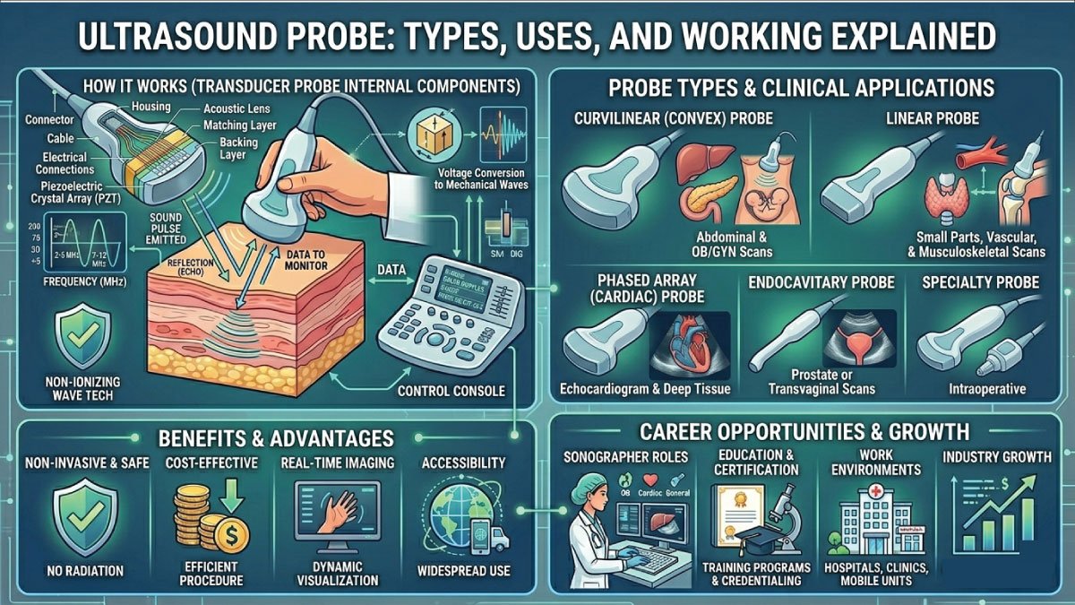
What Is an Ultrasound Probe?
An ultrasound probe, also known as an ultrasound transducer, is a handheld device used in diagnostic imaging to send and receive sound waves. These sound waves are beyond the range of human hearing (typically above 20 kHz), which is why they are called “ultrasound.”
The probe converts electrical energy into sound waves and then captures the echoes that bounce back from internal body structures. These echoes are transformed into images that appear on a monitor, helping doctors evaluate organs, tissues, and blood flow.
Key Components of an Ultrasound Probe:
- Piezoelectric crystals – Generate and receive sound waves
- Transducer housing – Protects internal components
- Cable and connector – Links the probe to the ultrasound machine
- Acoustic lens – Helps focus the ultrasound beam
How Does an Ultrasound Probe Work?
The ultrasound probe works using the piezoelectric effect, where crystals inside the probe convert electrical energy into sound waves and back into electrical signals. These waves travel into the body, reflect off tissues, and return as echoes, which are then transformed into real-time images for medical diagnosis.
Step-by-Step Process
1- Electrical Signal Generation
The process begins when the ultrasound machine generates an electrical signal and sends it to the probe. This signal is carefully controlled in terms of frequency and intensity, ensuring the probe can produce accurate sound waves needed for clear imaging of internal body structures.
2- Sound Wave Production
Inside the probe, piezoelectric crystals respond to the electrical signal by vibrating rapidly. These vibrations produce high-frequency sound waves that are transmitted into the body. This step is essential because it transforms electrical energy into mechanical energy used for imaging purposes.
3- Wave Transmission into the Body
The produced sound waves travel through the body and interact with different tissues. Some waves pass through easily, while others reflect or scatter depending on the density of the tissue. These interactions help create detailed differences in the final ultrasound image.
4- Echo Reception
When sound waves hit internal structures, they bounce back as echoes. The probe detects these returning echoes using the same piezoelectric crystals, which convert the sound energy back into electrical signals. This step is crucial for capturing information about internal organs.
5- Image Formation
The ultrasound machine processes the received electrical signals and calculates the time and strength of the echoes. Using this data, it creates a real-time visual image on the screen, allowing doctors to view organs, detect abnormalities, and monitor movement inside the body accurately.
Types of Ultrasound Probes
Different medical applications require different types of ultrasound probes. Each probe is designed with specific frequencies, shapes, and imaging capabilities to target particular organs or tissues. These variations allow healthcare professionals to obtain the clearest and most accurate images depending on the depth and type of examination required in clinical practice.
1. Linear Probe
A linear probe is designed to produce high-frequency sound waves that deliver very detailed and high-resolution images. It is mainly used for scanning shallow structures close to the skin surface. Because of its high clarity, it is ideal for examining small, superficial organs and detecting fine details in soft tissues.
Uses:
- Blood vessels
- Muscles and tendons
- Thyroid gland
- Breast imaging
Features:
- Flat surface
- High image clarity
- Limited penetration depth
2. Convex (Curvilinear) Probe
The convex probe has a curved surface that allows sound waves to spread out, creating a wider field of view. It is typically used for deeper imaging where greater penetration is needed. This makes it especially useful for examining internal organs located deep inside the abdomen or pelvis.
Uses:
- Abdominal scans
- Obstetrics and pregnancy
- Liver, kidneys, and spleen
Features:
- Lower frequency
- Greater penetration depth
- Wider field of view
3. Phased Array Probe
A phased array probe is specifically designed for imaging through small spaces, such as between the ribs. It uses electronically steered beams to capture detailed images of moving organs, especially the heart. This probe is widely used in cardiology because it provides real-time visualization of cardiac function and blood flow.
Uses:
- Cardiac imaging (echocardiography)
- Heart function analysis
Features:
- Small footprint
- Can scan between narrow spaces
- Ideal for dynamic organs
4. Endocavitary Probe
The endocavitary probe is designed for internal imaging by being inserted into body cavities. It provides highly detailed and close-range images, making it extremely useful in gynecological and urological examinations. Because of its proximity to internal organs, it offers better clarity and accuracy for detecting abnormalities.
Uses:
- Transvaginal ultrasound
- Transrectal ultrasound
- Prostate and pelvic examinations
Features:
- Inserted into body cavities
- Provides detailed internal images
- High-frequency for better resolution
5. Doppler Probe
A Doppler probe uses the Doppler effect to measure the movement and speed of blood flow within the body. It is an essential tool for evaluating circulation and detecting abnormalities in blood vessels. This probe helps doctors understand how blood flows through arteries and veins, making it vital in vascular and fetal monitoring.
Uses:
- Blood flow assessment
- Detecting blockages in arteries
- Monitoring fetal heartbeat
Features:
- Detects movement
- Measures velocity of blood flow
- Common in vascular studies
Also Read:
Uses of Ultrasound Probes in Medicine
Ultrasound probes are widely used in modern healthcare because they provide safe, real-time, and accurate imaging of internal body structures. Their versatility allows doctors to diagnose, monitor, and guide treatments across many medical specialties without using harmful radiation.
1. Obstetrics and Gynecology
One of the most important uses of ultrasound probes is in obstetrics and gynecology, especially during pregnancy. It allows doctors to monitor fetal growth, check the baby’s heartbeat, and detect any abnormalities. This helps ensure both mother and baby remain healthy throughout the pregnancy.
Uses:
- Track fetal development
- Check heartbeat
- Detect abnormalities
- Determine pregnancy stage
This non-invasive method is crucial for ensuring the health of both mother and baby.
2. Cardiology
In cardiology, ultrasound probes are used in a diagnostic test called echocardiography. This allows doctors to examine the heart in detail, evaluate its structure, and monitor how blood flows through it. It plays a vital role in detecting heart diseases and assessing overall heart function.
Uses:
- Evaluate heart structure
- Measure blood flow
- Detect heart diseases
- Assess valve function
3. Radiology and General Imaging
Ultrasound probes are commonly used in radiology to examine internal organs without surgery. They help visualize organs like the liver, kidneys, gallbladder, and pancreas. This makes it easier to detect conditions such as tumors, infections, or stones in a safe and non-invasive way.
Uses:
- Liver
- Kidneys
- Gallbladder
- Pancreas
This helps detect tumors, stones, and other abnormalities without surgery.
4. Musculoskeletal Imaging
Doctors use ultrasound probes to examine muscles, ligaments, tendons, and joints. This is especially useful for diagnosing sports injuries, sprains, and tears. It also helps guide injections and monitor healing progress in soft tissue injuries.
Uses:
- Muscles
- Ligaments
- Tendons
- Joints
This is useful for diagnosing injuries like sprains and tears.
5. Emergency Medicine
In emergency medicine, ultrasound probes are used for quick and life-saving assessments. They help detect internal bleeding, organ damage, and fluid accumulation in critical situations. This rapid imaging allows doctors to make immediate decisions and provide urgent treatment when time is very important.
Uses:
- Internal bleeding
- Organ damage
- Fluid accumulation
This rapid diagnosis can save lives in critical situations.
Advantages of Ultrasound Probes
Ultrasound probes offer several advantages over other imaging methods, making them a preferred choice in many medical situations. They are safe, efficient, and provide real-time imaging, which is essential for quick diagnosis and treatment.
1. Safe and Radiation-Free
Ultrasound does not use ionizing radiation, unlike X-rays or CT scans. This makes it safe for repeated use, especially for sensitive patients such as pregnant women and children. Doctors can perform multiple scans without health risks, ensuring safer diagnostic procedures.
2. Real-Time Imaging
Ultrasound provides real-time images, allowing doctors to observe movement inside the body. This is especially useful for monitoring heart activity and fetal development. It also helps during procedures where live imaging is needed for accuracy and precision.
3. Non-Invasive
Most ultrasound procedures are non-invasive and painless. There are no incisions or surgical tools involved, which reduces discomfort and recovery time. Patients can undergo the procedure without anxiety, making it one of the most patient-friendly imaging techniques available.
4. Cost-Effective
Compared to MRI and CT scans, ultrasound is more affordable and accessible. This makes it widely available in hospitals, clinics, and even rural healthcare settings. Its lower cost allows more patients to benefit from diagnostic imaging without financial burden.
5. Portable Technology
Modern ultrasound probes are compact and portable. Some can even connect to smartphones or tablets, making them useful in emergency situations and remote areas. This portability allows healthcare providers to deliver fast and efficient care wherever needed.
Limitations of Ultrasound Probes
Despite its advantages, ultrasound also has some limitations that affect its effectiveness in certain situations. These limitations depend on the type of tissue being examined and the skill of the operator performing the scan.
- Limited image quality for deep tissues compared to MRI
- Difficulty imaging through bone or air
- Operator-dependent results (requires skilled technician)
- Less effective for very obese patients
Despite these limitations, ultrasound remains one of the most widely used diagnostic tools.
Also Read:
Future of Ultrasound Probe Technology
The future of ultrasound technology is promising, with continuous innovations improving image quality, portability, and diagnostic accuracy. Advancements are making ultrasound more powerful, accessible, and easier to use across all medical fields.
1- Artificial Intelligence Integration
Artificial intelligence is enhancing ultrasound imaging by improving image interpretation and reducing human error. AI can assist doctors in detecting abnormalities faster and with greater accuracy, making diagnosis more reliable and efficient in clinical practice.
2- Portable and Wireless Devices
Modern ultrasound probes are becoming smaller, wireless, and more connected. Many devices now work with smartphones and tablets, allowing healthcare professionals to perform scans anywhere. This improves accessibility, especially in remote and emergency settings.
3- 3D and 4D Imaging
Advanced ultrasound probes can now create 3D and 4D images, providing more detailed visualization of internal structures. This is especially useful in obstetrics, where parents can see detailed images and real-time movement of the developing baby.
4- Improved Image Quality
Ongoing advancements in ultrasound technology are improving image resolution and clarity. Better imaging helps doctors detect diseases earlier and more accurately. These improvements continue to make ultrasound a more powerful and essential diagnostic tool in modern medicine.
Conclusion
The ultrasound probe is a vital tool in modern healthcare, offering safe, accurate, and real-time imaging of the human body. From pregnancy monitoring to diagnosing heart disease, its applications are vast and essential.
Understanding the different types of probes—such as linear, convex, phased array, and Doppler—helps appreciate how versatile this technology is. As advancements continue, ultrasound probes will become even more powerful, accessible, and intelligent.
If you’re exploring a career in medical imaging or simply curious about how diagnostic tools work, the ultrasound probe is a fascinating example of how science and technology come together to improve human health.
Also Read:

Zak is a dedicated medical and career writer specializing in sonography, healthcare education, and professional development. Through SonographerSalary.com, he shares in-depth insights on sonographer salaries, education pathways, and career tips to help readers build successful futures in medical imaging. His content combines accuracy with practical, easy-to-understand guidance, empowering students and professionals to make confident, informed career decisions.