
Ultrasound imaging is a cornerstone of modern medical diagnostics, offering a non-invasive and real-time view of organs, tissues, and blood flow. One of the most critical factors influencing image quality is the transducer frequency. Different frequencies affect resolution, depth penetration, and clinical application. Choosing the right frequency is crucial for accurate diagnosis and optimal patient care.
This guide explores ultrasound transducer frequencies, provides a frequency chart for all major applications, and explains how to select the correct transducer for every clinical scenario.
What Is Ultrasound Transducer Frequency?
Ultrasound transducer frequency determines how many sound wave cycles the probe emits per second, measured in megahertz (MHz). Choosing the right frequency is crucial for obtaining clear images, accurately visualizing tissues, and ensuring patient comfort. Different frequencies affect resolution, penetration depth, and overall diagnostic effectiveness.
Key points about frequency:
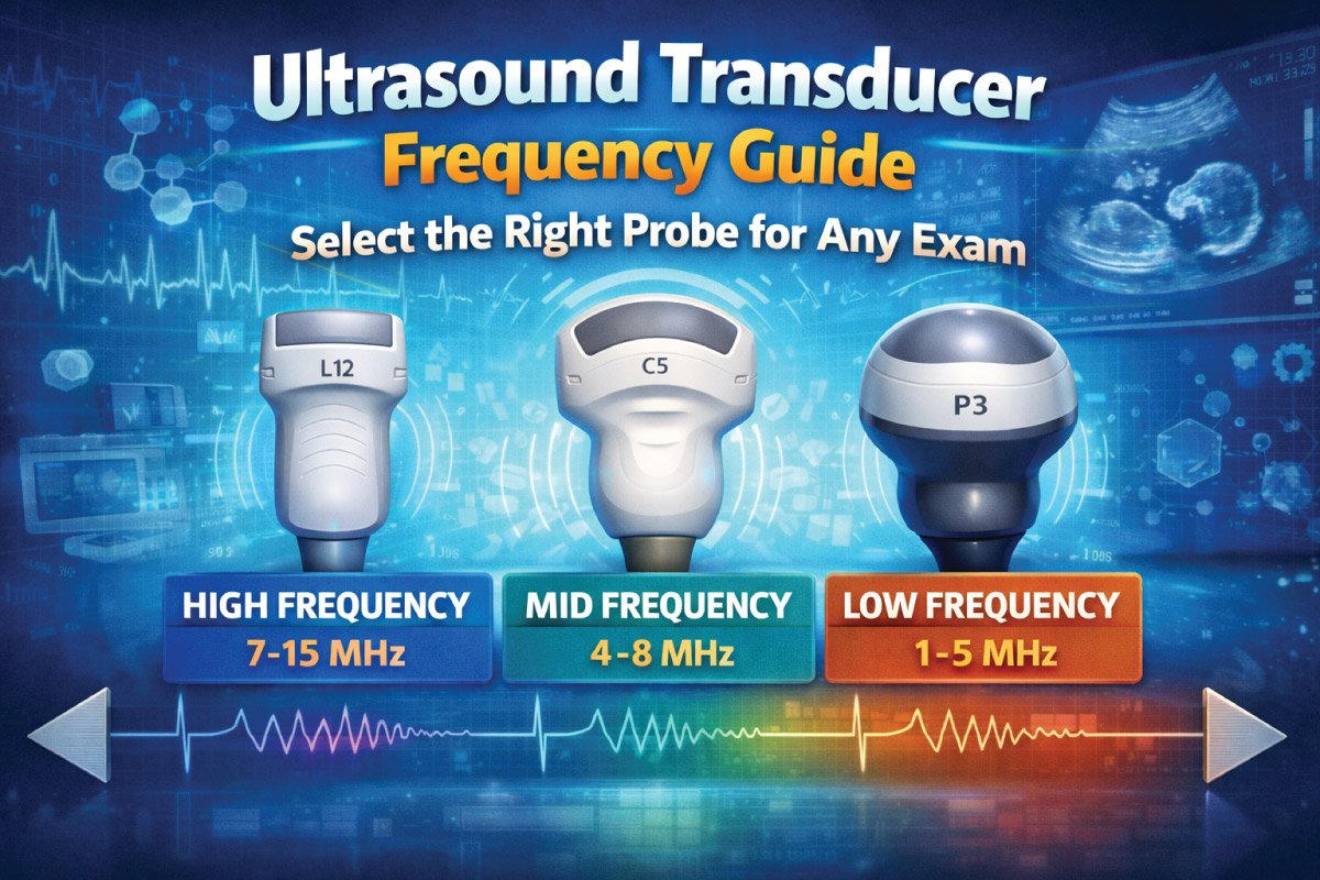
High-frequency transducers (10–18 MHz): Offer excellent image resolution but shallow penetration.
Low-frequency transducers (1–5 MHz): Penetrate deeper tissues but provide lower resolution images.
Frequency selection directly impacts diagnostic accuracy, scan time, and patient comfort.
How Frequency Affects Imaging
Ultrasound imaging relies on selecting the right transducer frequency, as it directly impacts image resolution and tissue penetration. Higher frequencies provide sharp, detailed images of superficial structures, while lower frequencies penetrate deeper but with moderate clarity. Understanding this trade-off is essential for accurate diagnosis across different clinical applications.
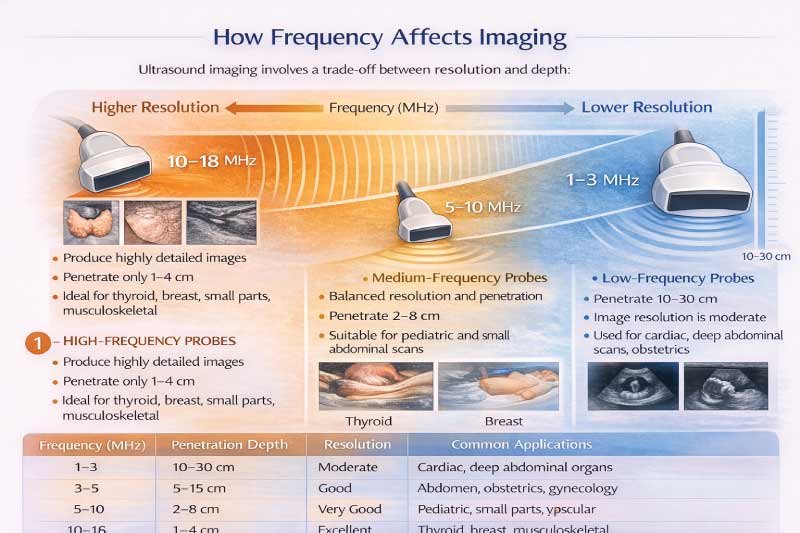
Ultrasound imaging involves a trade-off between resolution and depth:
1- High-Frequency Probes:
High-frequency probes produce exceptionally detailed images but have limited depth penetration (1–4 cm). They are ideal for imaging small, superficial structures such as the thyroid, breast, and musculoskeletal system, where precision and clarity are critical.
-
- Produce highly detailed images.
- Penetrate only 1–4 cm.
- Ideal for thyroid, breast, small parts, and musculoskeletal imaging.
2- Medium-Frequency Probes:
Medium-frequency probes strike a balance between image resolution and tissue penetration, reaching depths of 2–8 cm. They are well-suited for pediatric scans and small abdominal imaging, providing sufficient detail without sacrificing reach.
-
- Balanced resolution and penetration.
- Penetrate 2–8 cm.
- Suitable for pediatric and small abdominal scans.
3- Low-Frequency Probes:
Low-frequency probes penetrate deeper tissues (10–30 cm) but provide moderate resolution. They are essential for imaging larger or deep organs, including cardiac structures, adult abdominal organs, and obstetric assessments.
-
- Penetrate 10–30 cm.
- Image resolution is moderate.
- Used for cardiac imaging, deep abdominal scans, and obstetrics.
| Frequency (MHz) | Penetration Depth | Resolution | Common Applications |
|---|---|---|---|
| 1–3 | 10–30 cm | Moderate | Cardiac, deep abdominal organs |
| 3–5 | 5–15 cm | Good | Abdomen, obstetrics, gynecology |
| 5–10 | 2–8 cm | Very Good | Pediatric, small parts, vascular |
| 10–18 | 1–4 cm | Excellent | Thyroid, breast, musculoskeletal |
Frequency Chart for Common Ultrasound Applications
Ultrasound transducer frequency varies depending on the area being examined. Selecting the correct frequency ensures optimal image quality, accurate diagnosis, and efficient scanning. Higher frequencies provide better resolution for superficial structures, while lower frequencies penetrate deeper tissues for abdominal and cardiac imaging.
| Clinical Application | Recommended Frequency (MHz) | Recommended Probe Type | Depth Range | Notes |
|---|---|---|---|---|
| Abdominal (adult) | 2–5 | Convex | 10–20 cm | Liver, kidneys, pancreas |
| Obstetrics | 2–5 | Convex | 15–25 cm | Fetal imaging, placenta evaluation |
| Cardiac | 1–5 | Phased Array | 12–25 cm | Echocardiography, heart chambers |
| Vascular (arteries & veins) | 7–15 | Linear | 2–6 cm | Flow assessment, plaque detection |
| Thyroid | 10–18 | Linear | 1–4 cm | Nodules, cysts, small lesions |
| Breast | 10–15 | Linear | 2–4 cm | Mass detection, cyst evaluation |
| Musculoskeletal | 10–18 | Linear | 1–5 cm | Tendons, ligaments, joints |
| Pediatric Abdomen | 5–12 | Microconvex / Linear | 2–8 cm | Neonatal and infant organs |
| Transvaginal | 5–9 | Endocavitary | 5–10 cm | Early pregnancy, gynecology |
Pediatric and Neonatal Frequency Chart
Pediatric ultrasound requires smaller probes and carefully chosen frequencies to capture detailed images of tiny organs. Higher frequencies provide superior resolution for superficial structures, while lower frequencies are used for deeper tissues. Proper frequency selection ensures accurate diagnosis and patient safety in neonates, infants, and children.
| Age Group | Recommended Frequency (MHz) | Probe Type | Depth | Notes |
|---|---|---|---|---|
| Neonates (0–1 month) | 7–12 | Microconvex | 2–6 cm | Head, abdomen, heart |
| Infants (1–12 months) | 5–12 | Linear / Microconvex | 2–8 cm | Abdomen and small organs |
| Toddlers (1–3 years) | 5–10 | Convex / Linear | 5–10 cm | Abdomen, soft tissues |
| Children (3–12 years) | 3–7 | Convex | 5–15 cm | Standard abdominal scans |
Tip: Use the highest frequency possible for superficial structures, switching to a lower frequency for deeper organs.
Frequency Selection for Obstetric Imaging
In obstetric ultrasound, choosing the correct transducer frequency is essential for capturing clear images at each stage of pregnancy. Higher frequencies provide detailed views of early fetal structures, while lower frequencies are needed later for imaging larger fetuses and deeper maternal organs, ensuring accurate assessment and safe monitoring.
| Trimester | Frequency (MHz) | Probe Type | Depth | Notes |
|---|---|---|---|---|
| 1st Trimester | 5–9 | Endocavitary / Convex | 5–10 cm | High-resolution early fetal imaging |
| 2nd Trimester | 3–5 | Convex | 10–20 cm | Fetal anatomy scan, placenta |
| 3rd Trimester | 2–5 | Convex | 15–25 cm | Larger fetus, deeper organs |
Choosing the Right Frequency: Practical Guidelines
Selecting the proper ultrasound frequency is essential for clear imaging, accurate diagnosis, and patient comfort. Probe choice depends on tissue depth, patient size, and target anatomy. Using the appropriate high, medium, or low-frequency probe ensures optimal resolution and penetration, reduces scan time, and improves overall imaging efficiency in clinical practice.
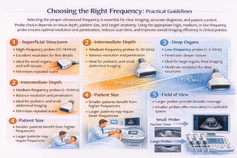
1- Superficial Structures:
Imaging the thyroid, breast, and musculoskeletal system requires high-frequency probes (10–18 MHz). These probes provide excellent resolution for fine details in superficial tissues, allowing precise evaluation of small organs, nodules, or soft tissue structures while minimizing the need for repeated scans.
2- Intermediate Depth:
Pediatric and small abdominal imaging are best performed with medium-frequency probes (5–10 MHz). These frequencies balance sufficient tissue penetration with good resolution, making them ideal for visualizing small organs, pediatric anatomy, and soft tissue structures without compromising image clarity.
3- Deep Organs:
Adult abdominal scans, cardiac imaging, and late-term obstetric examinations require low-frequency probes (1–5 MHz). These probes penetrate deeper tissues, allowing visualization of large organs, fetal structures, and deep vessels, although image resolution is moderate compared to higher-frequency probes.
4- Patient Size:
Smaller patients, such as children or neonates, allow higher-frequency imaging for improved resolution. Larger patients may require lower-frequency probes to achieve sufficient penetration. Proper frequency selection ensures clear images regardless of body habitus and maintains diagnostic accuracy.
5- Field of View:
Larger probes can provide a wider coverage area, which is useful for scanning broad regions. However, they may reduce detail in smaller, confined spaces. Choosing the right probe size ensures the best compromise between coverage and image detail for each clinical application.
Advanced Frequency Considerations
Modern ultrasound imaging offers advanced techniques that require careful frequency selection to optimize image quality and diagnostic accuracy. Harmonic, Doppler, and 3D/4D imaging each rely on specific frequencies to enhance resolution, capture flow dynamics, or generate volumetric images, enabling clinicians to visualize structures and functions more precisely than conventional imaging.
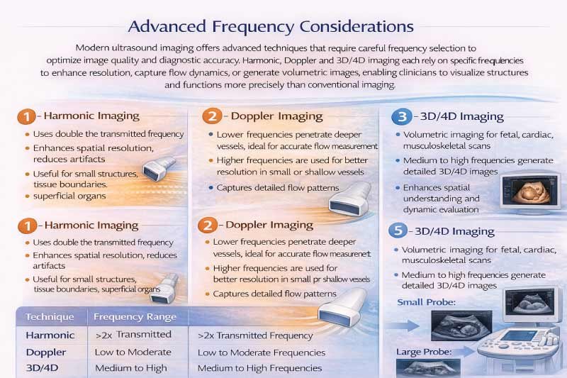
1- Harmonic Imaging:
This technique improves image clarity by using twice the transmitted frequency, which enhances spatial resolution and reduces artifacts. It is particularly useful for visualizing small structures, tissue boundaries, and superficial organs while maintaining image quality in challenging patients or areas.
2- Doppler Imaging:
Blood flow assessment depends on probe frequency. Lower frequencies are used for deeper vessels, allowing penetration and accurate flow measurement, while higher frequencies are ideal for superficial vessels, providing better resolution and detailed visualization of flow patterns in small or shallow vessels.
3- 3D/4D Imaging:
Volumetric imaging for fetal, cardiac, or musculoskeletal scans typically uses medium to high-frequency probes. This approach allows the generation of detailed three-dimensional or real-time four-dimensional images, enhancing spatial understanding, structural assessment, and dynamic evaluation for advanced diagnostic applications.
Also Read:
Frequency vs. Depth Chart Summary
Selecting the correct ultrasound frequency is key for balancing image quality and tissue penetration. Higher frequencies give excellent resolution for superficial structures, while lower frequencies penetrate deeper tissues. Proper frequency choice ensures accurate imaging, faster scans, and optimal diagnostic results across different clinical applications.
| Frequency (MHz) | Typical Depth (cm) | Image Quality | Ideal Use |
|---|---|---|---|
| 1–3 | 10–30 | Moderate | Cardiac, deep abdomen, large adults |
| 3–5 | 5–15 | Good | Abdominal, obstetrics, gynecology |
| 5–10 | 2–8 | Very Good | Pediatrics, small parts, vascular |
| 10–18 | 1–4 | Excellent | Thyroid, breast, musculoskeletal |
Pro Tip: Always start with a higher frequency probe when possible and switch to a lower frequency if the target is too deep.
Future of Ultrasound Frequencies
Advances in ultrasound technology are transforming how frequencies are used. Modern machines allow dynamic adjustments, improving image quality and workflow. Clinicians can now optimize resolution and penetration in real time, use versatile broadband transducers, and leverage AI-assisted frequency selection, enhancing diagnostic accuracy and patient care.
Optimize resolution and penetration without changing probes
Use broadband transducers covering multiple frequencies
Integrate AI algorithms for automatic frequency adjustment
These advancements enhance image quality, workflow efficiency, and diagnostic accuracy.
Conclusion
Understanding ultrasound transducer frequency is critical for accurate imaging and reliable diagnosis. Selecting the right frequency balances resolution and penetration based on clinical application, patient size, and target organ depth.
Using frequency charts and guidelines helps clinicians choose the optimal probe for any scenario—from pediatric exams to cardiac and obstetric imaging—ensuring efficient, precise, and patient-friendly ultrasound studies across all clinical settings.
Also Read:
FAQs:
What is an ultrasound probe?
An ultrasound probe, also called a transducer, is a handheld device that emits sound waves and receives echoes to create images of internal body structures. It converts electrical energy into sound waves and back into electrical signals, enabling real-time imaging for diagnosis in medical settings.
Types of transducers for ultrasound?
Ultrasound transducers come in several types based on imaging needs: linear, convex (curved), phased array, endocavitary, and microconvex. Each type differs in shape, frequency, and field of view, allowing clinicians to visualize structures from superficial organs to deep abdominal or cardiac regions effectively.
How ultrasound transducer works?
An ultrasound transducer works by converting electrical signals into sound waves, which travel through tissues and reflect back as echoes. The probe receives these echoes and converts them into electrical signals, which the machine processes to create real-time images of internal organs, blood flow, or fetal structures.
Types of ultrasonic transducers?
Ultrasonic transducers can be categorized as linear, convex, phased array, endocavitary, and microconvex. They differ in frequency, field of view, and penetration depth. Higher frequencies are ideal for superficial imaging, while lower frequencies penetrate deeper tissues, enabling versatile clinical applications.
Ultrasound transducer parts and function?
Key parts include the piezoelectric crystal, which emits and receives sound waves; the casing, which protects components; the cable, transmitting signals; and the connector, linking to the ultrasound machine. Each part works together to generate, transmit, receive, and process sound waves into clear diagnostic images.
Ultrasound transducer meaning?
An ultrasound transducer is a device that emits sound waves into the body and captures the returning echoes to form images. It serves as the bridge between the ultrasound machine and the patient, converting electrical energy into mechanical waves and back for real-time imaging.

Zak is a dedicated medical and career writer specializing in sonography, healthcare education, and professional development. Through SonographerSalary.com, he shares in-depth insights on sonographer salaries, education pathways, and career tips to help readers build successful futures in medical imaging. His content combines accuracy with practical, easy-to-understand guidance, empowering students and professionals to make confident, informed career decisions.