
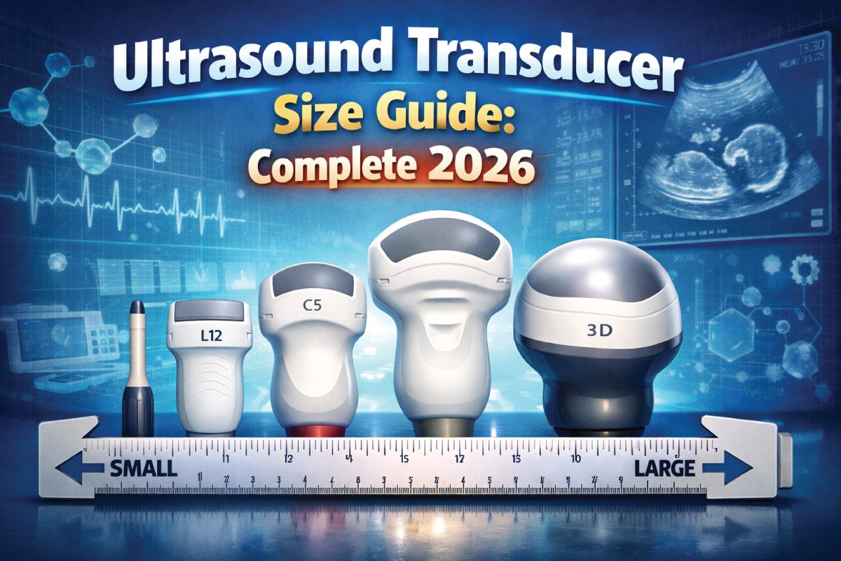
Ultrasound imaging is an essential diagnostic tool used across radiology, obstetrics, cardiology, and emergency medicine. The accuracy of an ultrasound scan largely depends on the transducer, also called a probe. Selecting the right transducer size and type ensures clear imaging, faster exams, and patient comfort.
This guide provides a complete overview of ultrasound transducer sizes, types, frequencies, and applications, helping healthcare professionals choose the right probe for every clinical scenario.
What Is an Ultrasound Transducer?
An ultrasound transducer is a device that sends sound waves into the body and receives the returning echoes to create real-time images. It contains piezoelectric crystals that vibrate when electricity passes through them, producing ultrasound waves. The returning echoes are converted into electrical signals that form images on the machine screen.
Key Functions:
- Emitting sound waves at specific frequencies
- Receiving echoes from tissue boundaries
- Producing diagnostic-quality images
How Ultrasound Transducer Size Affects Imaging
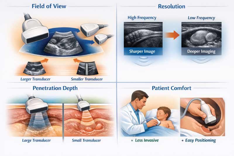
Ultrasound transducer size plays a critical role in image quality, depth, and patient comfort. Choosing the correct probe size ensures accurate visualization of organs and tissues while improving examination efficiency. The size of a transducer directly influences field of view, resolution, penetration depth, and overall patient experience during scanning.
1- Field of View (FOV)
Transducer size affects the field of view during an ultrasound exam. Larger transducers provide a wider viewing area, making them ideal for abdominal and obstetric imaging. Smaller transducers offer a narrower field of view but allow access to tight anatomical spaces such as intercostal regions.
2- Resolution
Smaller ultrasound probes typically operate at higher frequencies, which improves image resolution for superficial structures like tendons, vessels, and thyroid tissue. While higher resolution produces sharper detail, it comes at the cost of reduced penetration, limiting their use in deeper imaging applications.
3- Penetration Depth
Larger transducers usually operate at lower frequencies, allowing sound waves to penetrate deeper into the body. This makes them suitable for imaging deep organs such as the liver or kidneys. Smaller, high-frequency probes are less effective for deep penetration but excel in surface imaging.
4- Patient Comfort
Transducer size significantly impacts patient comfort during ultrasound examinations. Smaller probes are less invasive and easier to position, making them ideal for sensitive exams such as pediatric, vascular, or endo cavitary procedures. Improved comfort also helps reduce patient movement, enhancing image quality.
Types of Ultrasound Transducers
Ultrasound transducers come in different shapes, sizes, and frequency ranges to meet diverse clinical needs. Each transducer type is designed to optimize image quality for specific body areas and diagnostic purposes. Selecting the correct transducer improves accuracy, penetration, resolution, and overall efficiency during ultrasound examinations.
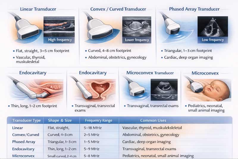
1- Linear Transducer
Linear transducers have a flat, straight footprint and operate at high frequencies, providing excellent resolution for superficial structures. They are commonly used for vascular imaging, thyroid exams, and musculoskeletal assessments where detailed visualization of shallow tissues is required.
2- Convex / Curved Transducer
Convex or curved transducers feature a wide, curved footprint that allows deeper penetration with a broader field of view. They operate at lower frequencies, making them ideal for abdominal, obstetric, and gynecological imaging, where deeper organs and fetal structures must be clearly visualized.
3- Phased Array Transducer
Phased array transducers have a small, triangular footprint that fits easily between ribs. They operate at low frequencies, allowing deep tissue penetration. These transducers are primarily used in cardiac imaging and deep organ studies where space is limited and depth is critical.
4- Endocavitary Transducer
Endocavitary transducers are thin, elongated probes designed for internal examinations. They provide high-resolution images at close range and are commonly used for transvaginal and transrectal exams. Their design allows detailed imaging of pelvic organs and nearby structures.
5- Microconvex Transducer
Microconvex transducers have a small curved footprint and operate at moderate frequencies. They are well-suited for pediatric, neonatal, and small-animal imaging, offering a balance between penetration and resolution while fitting comfortably on small anatomical areas.
| Transducer Type | Shape & Size | Frequency Range | Common Uses |
|---|---|---|---|
| Linear | Flat, straight, 3–5 cm footprint | 5–18 MHz | Vascular, thyroid, musculoskeletal |
| Convex / Curved | Curved, 4–8 cm footprint | 2–5 MHz | Abdominal, obstetrics, gynecology |
| Phased Array | Triangular, 1–3 cm footprint | 1–5 MHz | Cardiac, deep organ imaging |
| Endocavitary | Thin, long, 1–2 cm footprint | 5–9 MHz | Transvaginal, transrectal exams |
| Microconvex | Small curved, 2–4 cm footprint | 5–8 MHz | Pediatrics, neonatal, small animal imaging |
Understanding Frequency vs. Size
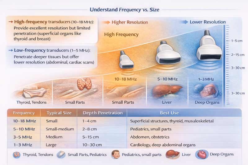
Ultrasound transducer frequency affects image resolution and tissue penetration. Higher frequencies produce sharper images but cannot reach deep structures, while lower frequencies penetrate deeper but with reduced detail. Understanding the relationship between frequency and probe size helps clinicians select the optimal transducer for accurate imaging in different clinical scenarios.
High-frequency transducers (10–18 MHz): Provide excellent resolution but limited penetration (superficial organs like thyroid and breast).
Low-frequency transducers (1–5 MHz): Penetrate deeper tissues but offer lower resolution (abdominal, cardiac scans).
| Frequency | Typical Size | Depth Penetration | Best Use |
|---|---|---|---|
| 10–18 MHz | Small | 1–4 cm | Superficial structures, thyroid, musculoskeletal |
| 5–10 MHz | Small-medium | 2–8 cm | Pediatrics, small parts |
| 3–5 MHz | Medium | 5–15 cm | Abdomen, obstetrics |
| 1–3 MHz | Large | 10–30 cm | Cardiology, deep abdominal organs |
Ultrasound Transducer Size Guide by Clinical Application
Selecting the correct ultrasound transducer for each clinical application ensures optimal imaging quality, accurate diagnosis, and patient safety. Different organs and examination types require specific probe sizes, footprints, and frequencies. Matching the transducer to the clinical need improves resolution, penetration depth, and overall exam efficiency.
| Clinical Application | Recommended Transducer Type | Size / Footprint | Frequency | Notes |
|---|---|---|---|---|
| Abdominal | Convex | 4–8 cm | 2–5 MHz | Liver, kidneys, pancreas |
| Obstetrics | Convex | 4–8 cm | 2–5 MHz | Fetal monitoring, placental imaging |
| Cardiac | Phased Array | 1–3 cm | 1–5 MHz | Heart imaging, echocardiography |
| Vascular | Linear | 3–5 cm | 7–15 MHz | Arteries and veins |
| Thyroid | Linear | 3–4 cm | 10–18 MHz | Nodules, masses, small structures |
| Musculoskeletal | Linear | 3–5 cm | 10–18 MHz | Tendons, ligaments, superficial joints |
| Transvaginal | Endocavitary | 1–2 cm | 5–9 MHz | Early pregnancy, gynecology |
| Pediatrics / Neonatal | Microconvex | 2–4 cm | 5–8 MHz | Small organs, neonatal scans |
Pediatric Ultrasound Transducer Size Guide
Pediatric and neonatal ultrasound imaging requires smaller, high-frequency probes to provide accurate images while ensuring patient comfort. Probe selection depends on the child’s age, size, and the target organ. Using the correct transducer improves diagnostic precision and minimizes discomfort during sensitive or detailed examinations.
| Age Group | Recommended Probe | Footprint | Frequency | Application |
|---|---|---|---|---|
| Neonates (0–1 month) | Microconvex | 2–3 cm | 7–12 MHz | Head, abdomen, heart |
| Infants (1–12 months) | Linear / Microconvex | 3–4 cm | 5–12 MHz | Abdomen, soft tissues |
| Toddlers (1–3 years) | Convex / Linear | 4–5 cm | 5–10 MHz | Abdominal scans |
| Children (3–12 years) | Convex | 5–8 cm | 3–7 MHz | Standard abdominal, organ evaluation |
Tip: Always start with the highest frequency probe suitable for the organ, switching to a lower frequency for deeper structures.
Obstetric Ultrasound Transducer Sizes
Obstetric ultrasound imaging requires different transducer sizes and frequencies as pregnancy progresses. Changes in fetal size, depth, and imaging goals across trimesters influence probe selection. Using the appropriate transducer improves image clarity, diagnostic accuracy, and patient comfort throughout all stages of pregnancy monitoring.
| Trimester | Recommended Probe | Footprint | Frequency | Notes |
|---|---|---|---|---|
| 1st Trimester | Endocavitary / Convex | 1–4 cm | 5–9 MHz | Early fetal development, high-resolution imaging |
| 2nd Trimester | Convex | 4–8 cm | 3–5 MHz | Fetal anatomy scans, amniotic fluid evaluation |
| 3rd Trimester | Convex | 4–8 cm | 2–5 MHz | Deeper fetal imaging, placenta, and growth monitoring |
Probe Footprint and Clinical Considerations
The footprint of an ultrasound probe directly affects image access, field of view, and patient comfort. Selecting the correct footprint ensures better visualization of target anatomy, especially in restricted spaces or sensitive examinations. Understanding footprint differences helps clinicians optimize imaging accuracy while improving patient experience across various clinical settings.
Small footprint: Ideal for narrow spaces (intercostal cardiac scans, neonatal imaging).
Large footprint: Wider field of view for large organs (liver, kidney).
Patient comfort: Smaller probes reduce discomfort in transvaginal and pediatric exams.
| Probe Type | Footprint (cm) | Field of View | Typical Use |
|---|---|---|---|
| Linear | 3–5 | Wide, shallow | Vascular, thyroid, musculoskeletal |
| Convex | 4–8 | Moderate | Abdominal, obstetrics |
| Phased Array | 1–3 | Narrow, deep | Cardiac |
| Microconvex | 2–4 | Small, curved | Pediatric, neonatal |
| Endocavitary | 1–2 | Narrow, close | Vaginal/rectal exams |
Factors to Consider When Choosing a Transducer
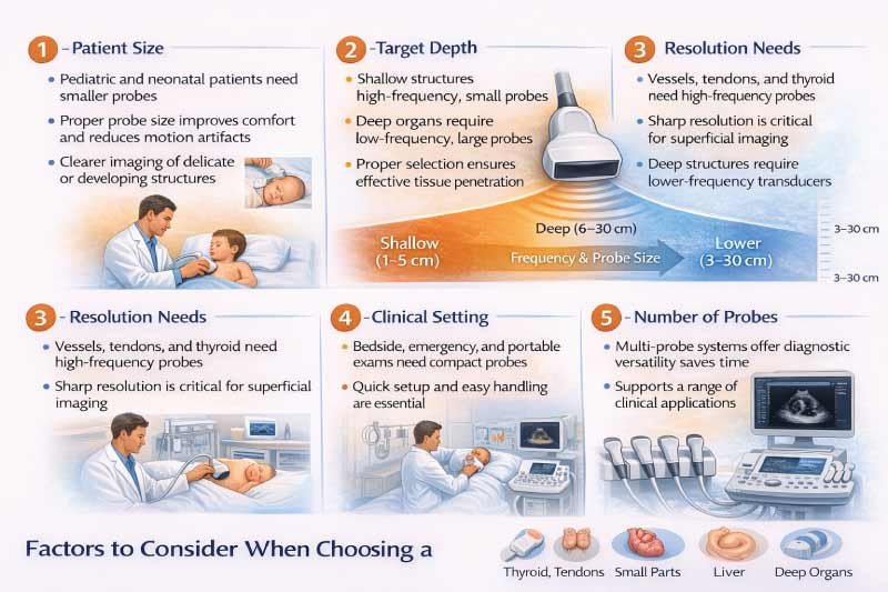
Selecting the right ultrasound transducer is essential for accurate imaging and efficient examinations. Factors such as patient size, target depth, resolution requirements, and clinical environment all influence probe selection. Choosing the correct transducer improves image quality, examination comfort, and diagnostic confidence across different medical applications.
1- Patient Size
Patient size plays a major role in transducer selection. Pediatric and neonatal patients require smaller probes that fit comfortably on smaller anatomical areas. Using appropriately sized transducers improves patient comfort, reduces motion artifacts, and ensures clearer imaging of delicate or developing structures.
2- Target Depth
The depth of the target organ determines the frequency and size of the transducer. Deeper organs require low-frequency, larger probes to allow sound waves to penetrate tissues effectively. High-frequency probes may produce clearer images but cannot adequately reach deep anatomical structures.
3- Resolution Needs
Resolution requirements vary based on the structure being examined. Superficial tissues such as vessels, tendons, and thyroid glands require high-frequency transducers for detailed imaging. These probes provide sharp resolution but are limited in penetration, making them unsuitable for deep imaging.
4- Clinical Setting
The clinical environment influences transducer choice. Bedside, emergency, or portable exams often benefit from smaller or microconvex probes due to ease of handling and quick setup. Compact probes enhance mobility and flexibility while maintaining acceptable image quality in fast-paced settings.
5- Number of Probes
The number of available transducers affects diagnostic versatility. Multi-probe systems allow clinicians to switch between different probe types for various exams without changing machines. This flexibility improves workflow efficiency and supports a wider range of clinical applications.
Tips for Maintaining Ultrasound Transducers
Proper maintenance of ultrasound transducers is essential for preserving image quality, patient safety, and equipment longevity. Routine cleaning, careful handling, and correct storage help prevent damage and reduce the risk of cross-contamination, ensuring reliable performance during every ultrasound examination.
-
Clean after every use with approved disinfectants
-
Inspect cables and crystals for damage
-
Store properly in holders to prevent wear
-
Avoid dropping or excessive bending
Proper care prolongs probe life and ensures consistent image quality.
Future Trends in Ultrasound Transducer Sizes
Ultrasound transducer technology is rapidly evolving to meet the demands of modern healthcare. Advances in miniaturization, wireless design, and artificial intelligence are improving portability, image quality, and ease of use. These innovations allow clinicians to perform faster, more accurate imaging across diverse clinical environments.
-
Miniaturized probes: More portable and lightweight
-
Wireless connectivity: Enables point-of-care imaging without cables
-
AI-assisted imaging: Auto-optimization of depth and resolution
-
3D/4D integration: High-end transducers for fetal and cardiac imaging
These advancements make transducers more versatile, user-friendly, and efficient for modern healthcare settings.
Also Read:
Conclusion
The size of an ultrasound transducer plays a vital role in obtaining accurate and efficient diagnostic images. Understanding how probe size, frequency, and penetration depth relate to specific clinical applications helps clinicians select the right transducer.
Whether for pediatric, obstetric, cardiac, or musculoskeletal imaging, the correct probe ensures superior image quality, faster exams, patient comfort, and precise diagnosis. Using this guide, healthcare professionals can optimize workflow and improve patient outcomes.
✅ Pro Tip: Always match the transducer size and frequency to the organ and patient type. Keep a reference chart for all transducer types to save time and optimize imaging quality.
FAQs:
What Is an Ultrasound Probe?
An ultrasound probe, also called a transducer, is a device that emits high-frequency sound waves into the body. It captures echoes reflected from tissues and organs and sends them to the ultrasound machine, which creates real-time images. Probes come in various shapes and sizes depending on the clinical application.
Types of Transducers for Ultrasound?
Ultrasound transducers vary in shape, size, and frequency. Common types include linear, convex (curved), phased array, endocavitary, and microconvex probes. Each type is designed for specific imaging needs, such as vascular studies, abdominal scans, cardiac imaging, obstetrics, or pediatric exams, to optimize resolution and penetration depth.
How Ultrasound Transducer Works?
An ultrasound transducer works by converting electrical energy into high-frequency sound waves. These waves travel into the body, reflect off tissues, and return to the probe as echoes. The ultrasound machine processes these echoes into real-time images, allowing clinicians to evaluate organs, blood flow, and other structures without radiation.
Types of Ultrasonic Transducers?
Ultrasonic transducers are classified based on their shape, frequency, and application. The main types are linear, convex, phased array, endocavitary, and microconvex. Each type has specific advantages, such as high resolution for superficial imaging, deep tissue penetration, or suitability for specialized exams like cardiac or obstetric imaging.
Ultrasound Transducer Definition?
An ultrasound transducer is a medical device that generates and receives sound waves to produce images of internal body structures. Also called a probe, it converts electrical signals into sound waves and back, allowing visualization of organs, blood vessels, and fetal development in real-time for diagnostic purposes.
Ultrasound Transducer Parts and Function?
Ultrasound transducers consist of a probe housing, piezoelectric crystals, cables, and connectors. The crystals generate and receive sound waves, the housing protects components, and cables transmit signals to the machine. Some probes have multiple crystals or arrays for different imaging modes, enhancing resolution, penetration, and clinical versatility.
Ultrasound Transducer Meaning?
An ultrasound transducer, or probe, is the part of the ultrasound system that contacts the patient’s body to send and receive sound waves. It enables the machine to capture echoes and create images, allowing clinicians to visualize tissues, organs, and blood flow safely, without radiation, in real-time for diagnosis and monitoring.

Zak is a dedicated medical and career writer specializing in sonography, healthcare education, and professional development. Through SonographerSalary.com, he shares in-depth insights on sonographer salaries, education pathways, and career tips to help readers build successful futures in medical imaging. His content combines accuracy with practical, easy-to-understand guidance, empowering students and professionals to make confident, informed career decisions.