
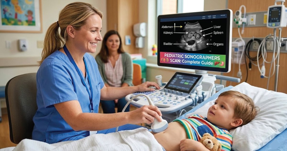
A pediatric sonographer is a highly trained medical professional who uses ultrasound technology to examine infants, children, and adolescents.
Unlike adult sonography, pediatric imaging requires specialized techniques, high-frequency probes, and a compassionate approach because children are smaller, developing rapidly, and often anxious during scans.
This guide explains everything pediatric sonographers scan, the purpose of each scan, the conditions they detect, and the special considerations involved.
1. Abdominal Ultrasound
Abdominal ultrasound is a core pediatric imaging study used to evaluate internal abdominal organs in infants and children. It helps detect structural abnormalities, infections, congenital conditions, and emergency issues quickly, safely, and without radiation exposure.
What It Is
Abdominal ultrasound is one of the most common scans performed by pediatric sonographers. It evaluates the organs and structures within the child’s abdomen using high-frequency sound waves.
Organs Scanned
-
Liver – Assess size, structure, and liver disease
-
Spleen – Evaluate for enlargement or injury
-
Kidneys – Check for congenital abnormalities, hydronephrosis, or obstruction
-
Gallbladder and bile ducts – Detect stones or inflammation
-
Pancreas – Examine for cysts, tumors, or inflammation
-
Bladder – Assess urinary issues
Conditions Detected
- Abdominal masses
- Hydronephrosis
- Appendicitis
- Congenital organ abnormalities
- Inflammatory conditions
Pediatric Considerations
- Children may move or cry, so quick imaging is crucial
- Requires adjusting transducer size and frequency for small organs
2. Neonatal Cranial (Brain) Ultrasound
Neonatal cranial ultrasound is a specialized brain scan performed in newborns, especially premature infants. It plays a vital role in early detection of brain injuries, bleeding, or developmental abnormalities without exposing fragile babies to radiation.
What It Is
Performed through the fontanelle (soft spot) in newborns, this ultrasound allows imaging of the brain without radiation.
Structures Scanned
- Brain ventricles
- Cerebral cortex
- White matter
- Subependymal areas
Conditions Detected
- Brain hemorrhage in premature infants
- Hydrocephalus (fluid accumulation in the brain)
- Periventricular leukomalacia (white matter injury)
- Congenital brain abnormalities
Pediatric Considerations
- Usually performed in the NICU
- Requires gentle handling and minimal movement
- Often repeated for monitoring progress
3. Hip Ultrasound
Hip ultrasound is primarily used in newborn screening to ensure proper hip joint development. Early detection of hip instability prevents long-term mobility problems and supports timely treatment during critical growth stages.
What It Is
Hip ultrasound is commonly performed on newborns to detect developmental dysplasia of the hip (DDH).
Structures Scanned
- Femoral head
- Acetabulum (hip socket)
- Ligaments and joint capsule
Conditions Detected
- Hip instability or dislocation
- Abnormal joint development
Pediatric Considerations
- Positioning is critical; sometimes special pillows or braces are used
- Quick and gentle scanning is necessary for newborn comfort
Discover More:
4. Renal and Urinary Ultrasound
Renal and urinary ultrasound helps evaluate kidney and bladder health in infants and children. It is commonly ordered for urinary infections, congenital kidney conditions, or unexplained abdominal symptoms.
What It Is
This scan evaluates the kidneys, ureters, and bladder in infants and children.
Structures Scanned
- Kidneys – Size, shape, echogenicity
- Bladder – Filling, emptying, structural abnormalities
- Ureters – Obstructions or dilation
Conditions Detected
- Congenital kidney malformations
- Hydronephrosis or obstruction
- Urinary tract infections (UTIs) complications
- Kidney stones
Pediatric Considerations
- May require bladder filling (child drinks water)
- Infants may need gentle restraint
Discover More:
5. Pelvic Ultrasound
Pelvic ultrasound in pediatric patients evaluates reproductive organs when hormonal, developmental, or emergency concerns arise. It is particularly important in assessing pain, masses, or congenital reproductive abnormalities.
What It Is
Used mainly in older infants and children to examine reproductive organs.
Structures Scanned
- Ovaries
- Uterus
- Testes
- Prostate (in male children if indicated)
Conditions Detected
- Ovarian cysts or masses
- Undescended testes
- Testicular torsion (emergency)
- Congenital anomalies of the reproductive tract
Pediatric Considerations
- Older children may need reassurance or distraction
- Scans are performed quickly to minimize anxiety
6. Vascular Ultrasound
Vascular ultrasound evaluates blood vessels and circulation in children. Using Doppler technology, it helps detect abnormalities in blood flow, vessel structure, and rare pediatric clotting conditions.
What It Is
This scan evaluates blood vessels and blood flow in babies and children using Doppler imaging.
Structures Scanned
- Arteries (carotid, femoral, renal)
- Veins (deep veins for thrombosis)
- Heart vessels (sometimes in echocardiography)
Conditions Detected
- Vascular malformations
- Blood clots (rare but possible in children)
- Poor blood flow to organs
Pediatric Considerations
- High-frequency linear probes are used
- Requires patience for small vessels and fast heart rates
Discover More:
7. Soft Tissue and Musculoskeletal Ultrasound
Musculoskeletal ultrasound is widely used in pediatrics to assess injuries, inflammation, or unexplained soft tissue lumps. It offers a fast and non-invasive alternative to more advanced imaging methods.
What It Is
This scan is used to examine muscles, tendons, and soft tissue masses in children.
Structures Scanned
- Muscle and tendon injuries
- Subcutaneous masses
- Ligaments and joints (sometimes for trauma)
Conditions Detected
- Soft tissue tumors or cysts
- Inflammatory conditions (like juvenile arthritis)
- Trauma injuries
Pediatric Considerations
- Children may move frequently; distraction is needed
- Scans are non-invasive and often used instead of MRI
8. Cardiac Ultrasound (Echocardiography) – Sometimes Included
Cardiac ultrasound, also known as echocardiography, evaluates heart structure and function in infants and children. It is essential for diagnosing congenital heart defects and monitoring cardiac performance.
Structures Scanned
- Heart chambers
- Heart valves
- Major vessels (aorta, pulmonary artery)
Conditions Detected
- Congenital heart defects
- Valve abnormalities
- Heart function evaluation
Pediatric Considerations
- Often done in NICU or pediatric cardiology
- Requires precise timing with heartbeats
Discover More:
Special Pediatric Considerations for All Ultrasounds
Patient Cooperation
-
- Infants and children often cry, move, or become anxious.
- Sonographers use toys, pacifiers, and parent involvement to calm the child.
Size and Development
-
- Pediatric organs are smaller and still developing.
- High-frequency probes and fine adjustments are used for detailed imaging.
Safety
-
- Ultrasound is safe, but minimal pressure and short scan times are preferred.
Repeatability
-
- Some conditions require repeated scans over time to monitor growth or progression.
Summary Table: What Pediatric Sonographers Scan
| Scan Type | Age Group | Main Structures | Conditions Detected |
|---|---|---|---|
| Abdominal Ultrasound | Infants & children | Liver, spleen, kidneys, bladder | Masses, infections, organ anomalies |
| Cranial Ultrasound (Neonatal) | Newborns | Brain ventricles, white matter | Hemorrhage, hydrocephalus |
| Hip Ultrasound | Newborns | Femoral head, acetabulum | Developmental dysplasia |
| Renal & Urinary Ultrasound | Infants & children | Kidneys, ureters, bladder | Obstruction, congenital anomalies |
| Pelvic Ultrasound | Infants & older children | Ovaries, uterus, testes | Cysts, torsion, congenital anomalies |
| Vascular Ultrasound | Infants & children | Arteries, veins | Malformations, clots |
| Musculoskeletal & Soft Tissue | Infants & children | Muscles, tendons, ligaments | Injuries, masses, inflammation |
| Echocardiography | Infants & children | Heart chambers, valves, vessels | Congenital defects, functional evaluation |
Conclusion
Pediatric sonographers scan a wide range of organs and systems, including the brain, abdomen, hips, kidneys, bladder, reproductive organs, blood vessels, soft tissues, and sometimes the heart. Each scan is tailored to the age, size, and condition of the child.
They use high-frequency ultrasound, gentle handling, and child-friendly techniques to ensure accurate imaging while keeping young patients safe and comfortable. Their work is vital for early diagnosis, treatment planning, and monitoring of pediatric conditions.
In short, pediatric sonographers are experts in imaging babies and children across multiple organ systems, using ultrasound to provide safe, non-invasive, and life-saving diagnostic information.

Zak is a dedicated medical and career writer specializing in sonography, healthcare education, and professional development. Through SonographerSalary.com, he shares in-depth insights on sonographer salaries, education pathways, and career tips to help readers build successful futures in medical imaging. His content combines accuracy with practical, easy-to-understand guidance, empowering students and professionals to make confident, informed career decisions.