
The ultrasound transducer is the heart of every ultrasound system. Without it, there would be no imaging, no diagnosis, and no real-time visualization of the human body. Whether used in obstetrics, cardiology, or abdominal imaging, the transducer plays a critical role in converting electrical energy into sound waves and then interpreting the returning echoes to create images.
In this detailed guide, we will explore how an ultrasound transducer works, its different types, and its wide range of applications in modern medicine. This article includes related concepts, medical terminology, and supporting topics to give you a complete understanding.
What Is an Ultrasound Transducer?
An ultrasound transducer is a medical device that converts electrical signals into high-frequency sound waves (ultrasound) and then converts the returning echoes back into electrical signals.
These sound waves are beyond the range of human hearing (typically above 20 kHz) and are safe for internal imaging because they do not involve radiation. The transducer is used in combination with an ultrasound machine to create real-time images of organs, tissues, and blood flow.
In simple terms, the transducer acts like both a speaker and a microphone:
- It emits sound waves into the body
- It receives echoes from internal structures
- It sends data to the ultrasound system to form images
This process is known as the pulse-echo principle, which is fundamental in ultrasound imaging.
How Does an Ultrasound Transducer Work?
The working principle of an ultrasound transducer is based on the piezoelectric effect, a scientific concept that allows certain crystals to convert electrical energy into mechanical (sound) energy and vice versa.
Inside the transducer, there are piezoelectric crystals. When an electrical current is applied, these crystals vibrate and produce sound waves. When these sound waves hit internal body structures, they reflect back as echoes.
The transducer then:
- Receives the reflected echoes
- Converts them back into electrical signals
- Sends them to the ultrasound machine
- The machine processes the signals into a visual image
This entire process happens in real time, allowing doctors to observe movement such as blood flow or fetal activity instantly.
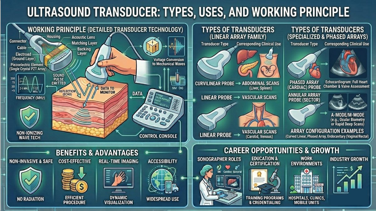
Key Components of a Transducer
- Piezoelectric crystals – Generate and receive sound waves
- Backing material – Absorbs unwanted vibrations
- Matching layer – Improves sound transmission into the body
- Lens – Focuses the sound waves
- Housing – Protects internal components
Types of Ultrasound Transducers
Ultrasound transducers come in several types, each designed for specific diagnostic purposes in medical imaging. Understanding their structure, frequency range, and application helps healthcare professionals choose the most suitable probe for accurate, high-quality imaging in different clinical scenarios.
1. Linear Transducer
The linear transducer is widely used for imaging superficial structures with high clarity and precision. It produces a rectangular image with excellent resolution, making it ideal for examining tissues near the skin surface and providing detailed diagnostic information.
- Frequency range: High (5–15 MHz)
- Penetration depth: Low
- Image quality: Excellent detail
Uses:
- Vascular imaging
- Musculoskeletal ultrasound
- Thyroid and breast imaging
- Superficial structures
This type is ideal for examining structures close to the skin because it provides detailed and sharp images.
2. Curvilinear (Convex) Transducer
The curvilinear transducer is designed with a curved surface that allows a wider field of view, making it highly effective for imaging deeper organs. Its lower frequency enables better penetration, though with slightly reduced image resolution compared to linear probes.
- Frequency range: Low (2–5 MHz)
- Penetration depth: High
- Image quality: Moderate
Uses:
- Abdominal scans
- Obstetrics (pregnancy imaging)
- Liver, kidneys, and pelvic organs
It is commonly used for deep internal organs because it can penetrate further into the body.
3. Phased Array Transducer
The phased array transducer is a compact and versatile probe that uses electronic beam steering to generate images. Its small footprint allows it to scan through narrow spaces, making it particularly useful in cardiac imaging and emergency applications.
- Frequency range: Low to medium
- Special feature: Small footprint
Uses:
- Cardiac imaging (echocardiography)
- Intercostal scanning (between ribs)
It is especially useful for imaging the heart because it can fit into narrow spaces.
4. Endocavitary Transducer
The endocavitary transducer is specifically designed for internal examinations by being inserted into body cavities. This allows for closer proximity to organs, resulting in highly detailed and accurate images for gynecological and urological assessments.
- Types:
- Transvaginal probe
- Transrectal probe
Uses:
- Gynecological exams
- Prostate imaging
- Early pregnancy assessment
This type provides highly detailed images of internal organs due to its close proximity.
5. Sector Transducer
The sector transducer produces a pie-shaped image and is often used in situations where space is limited. It is commonly associated with cardiac and neonatal imaging due to its ability to provide a wide view from a small contact area.
Uses:
- Cardiac imaging
- Neonatal brain imaging
It is useful in situations where access is limited.
Also Read:
Uses of Ultrasound Transducers in Medicine
Ultrasound transducers are essential tools in modern healthcare, used across a wide range of medical specialties. Their ability to produce real-time images without radiation makes them highly valuable for diagnosis, monitoring, and treatment planning in various clinical environments.
1. Obstetrics and Gynecology
Ultrasound transducers are essential in monitoring pregnancy and female reproductive health. They help doctors track fetal development and ensure the well-being of both mother and baby throughout different stages of pregnancy.
- Tracking fetal growth
- Checking heartbeat
- Detecting abnormalities
- Determining gestational age
This is one of the most common uses of ultrasound technology.
2. Cardiology
In cardiology, transducers are used in echocardiography to evaluate heart structure and function. They help in diagnosing cardiovascular conditions and monitoring heart performance with accurate, real-time imaging.
- Evaluating heart function
- Detecting valve disorders
- Measuring blood flow
- Identifying heart diseases
3. Abdominal Imaging
Ultrasound transducers are widely used to examine abdominal organs and detect abnormalities. They provide clear visualization of internal structures, helping doctors identify diseases or conditions affecting vital organs.
- Liver
- Kidneys
- Gallbladder
- Pancreas
They assist in detecting tumors, stones, or infections.
4. Musculoskeletal Imaging
This type of imaging focuses on muscles, tendons, ligaments, and joints. It is commonly used to diagnose injuries, monitor healing processes, and assess soft tissue conditions effectively.
- Diagnosing injuries
- Monitoring inflammation
- Evaluating soft tissue damage
5. Vascular Imaging
Ultrasound transducers play an important role in assessing blood vessels and circulation. They help detect vascular diseases and ensure proper blood flow throughout the body.
- Detecting blood clots
- Measuring blood flow
- Identifying blockages
6. Emergency Medicine
In emergency care, ultrasound transducers are used in the FAST exam to quickly assess trauma patients. This rapid imaging helps detect internal bleeding and guides immediate treatment decisions in critical situations.
Advantages of Ultrasound Transducers
Ultrasound transducers offer numerous advantages that make them a preferred choice in medical imaging. Their safety, efficiency, and accessibility contribute to their widespread use in hospitals and clinics around the world.
- Non-invasive – No surgery required
- Radiation-free – Safe for repeated use
- Real-time imaging – Immediate results
- Portable – Can be used in clinics or remote areas
- Cost-effective – More affordable than MRI or CT scans
These advantages make ultrasound one of the most widely used imaging techniques worldwide.
Limitations of Ultrasound Transducers
Despite their many benefits, ultrasound transducers also have certain limitations that affect their performance in specific situations. Understanding these limitations is important for accurate diagnosis and selecting the right imaging method.
- Limited penetration in obese patients
- Lower image quality compared to MRI
- Operator-dependent results
- Difficulty imaging air-filled organs (like lungs)
Understanding these limitations helps healthcare professionals choose the right imaging method.
Also Read:
Maintenance and Care of Transducers
Proper maintenance of ultrasound transducers is essential for ensuring accurate imaging and prolonging the life of the device. Regular cleaning and careful handling help maintain performance and prevent damage.
Best Practices:
- Clean and disinfect after each use
- Avoid dropping or damaging the probe
- Use proper gel during scanning
- Store in a safe, dry place
- Regularly inspect for wear and tear
Poor maintenance can lead to image distortion or device failure.
Future of Ultrasound Transducer Technology
The future of ultrasound transducers is rapidly evolving with advancements in imaging technology. New innovations are making ultrasound more precise, portable, and efficient in modern healthcare environments.
Key Innovations:
- 3D and 4D imaging – More detailed visualization
- Portable handheld devices – Increased accessibility
- AI integration – Improved diagnostic accuracy
- High-frequency probes – Better resolution
These innovations are making ultrasound faster, more accurate, and more accessible worldwide.
Conclusion
The ultrasound transducer is a powerful and essential tool in modern medicine. It works on the principle of converting electrical energy into sound waves and interpreting echoes to create real-time images of the human body.
From obstetrics to cardiology, and from emergency medicine to musculoskeletal imaging, transducers play a vital role in accurate diagnosis and patient care. With ongoing advancements in technology, ultrasound transducers are becoming even more precise, portable, and intelligent.
Understanding the types, uses, and working principles of ultrasound transducers helps medical professionals and students appreciate their importance in healthcare. As technology continues to evolve, these devices will remain at the forefront of non-invasive diagnostic imaging.
Also Read:

Zak is a dedicated medical and career writer specializing in sonography, healthcare education, and professional development. Through SonographerSalary.com, he shares in-depth insights on sonographer salaries, education pathways, and career tips to help readers build successful futures in medical imaging. His content combines accuracy with practical, easy-to-understand guidance, empowering students and professionals to make confident, informed career decisions.