
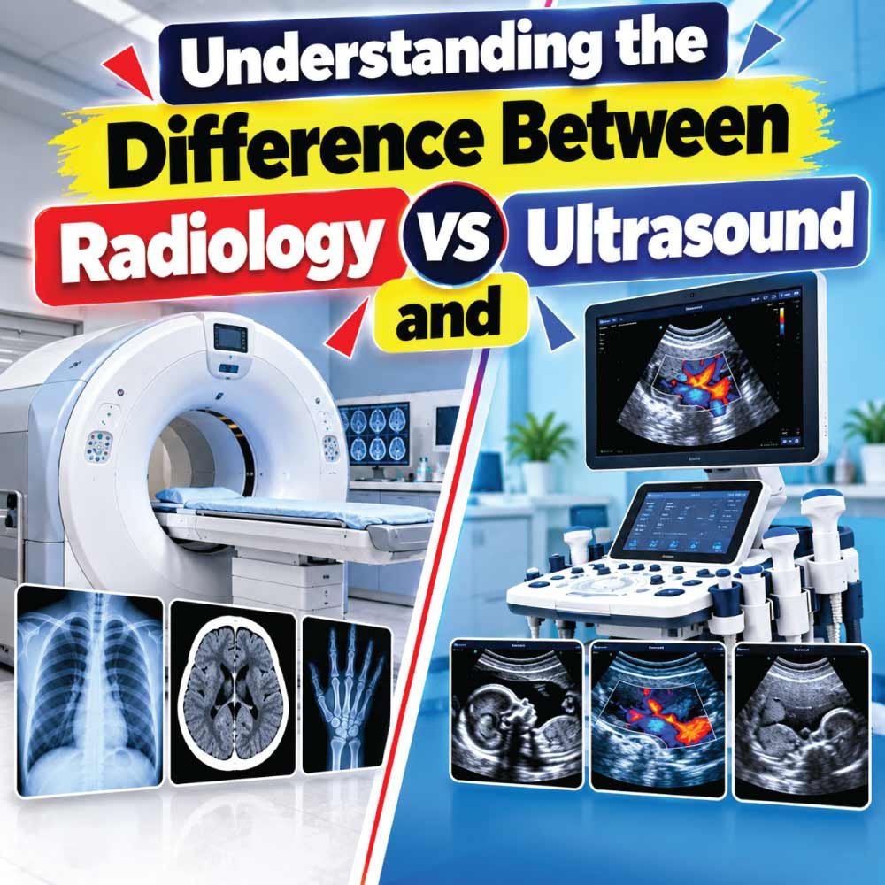
The difference between radiology and ultrasound can be confusing because both are used to create images of the inside of the body. However, they are not the same thing. Ultrasound, also called sonography, is actually one type of medical imaging that can be used within the broader field of radiology.
The main distinction is the technology involved. Ultrasound uses high-frequency sound waves and a handheld transducer to produce images. Many radiology examinations, by comparison, use technologies such as X-rays, computed tomography (CT), magnetic resonance imaging (MRI), or nuclear medicine.
Radiology is therefore a broad medical specialty, while ultrasound is a specific imaging method. The people who perform these examinations can also have different roles and training.
Understanding this distinction can help patients know what to expect from an examination and help students compare careers such as diagnostic medical sonography and radiologic technology.
What Is Radiology?
Radiology is a medical specialty focused on using imaging technology to examine the body and help diagnose or manage disease and injury. It encompasses several different imaging techniques rather than one single type of scan.
Depending on the clinical question, imaging may involve:
- X-rays
- CT scans
- MRI
- Ultrasound
- Nuclear medicine
- Fluoroscopy
- Interventional imaging procedures
A radiologist is a physician who specializes in interpreting medical images and, in some cases, performing image-guided procedures. Radiologists may work with images produced by X-rays, CT, MRI, ultrasound, and other modalities.
This means that saying someone is having a “radiology exam” does not necessarily mean they are receiving an X-ray. The specific examination determines the imaging technology used.
For example, a patient with a suspected fracture may receive an X-ray, while someone with certain abdominal symptoms may undergo an ultrasound. Another patient may require CT or MRI when more detailed information is needed.
What Is Ultrasound or Sonography?
Ultrasound imaging, commonly called sonography, is a medical imaging technique that uses high-frequency sound waves to create pictures of structures inside the body.
During an examination, a sonographer places a transducer against the patient’s skin, usually with a small amount of gel between the probe and the skin. The transducer sends sound waves into the body and receives the returning echoes. A computer processes those echoes and produces images on a monitor.
Unlike X-ray-based imaging, diagnostic ultrasound does not use ionizing radiation.
One important feature of ultrasound is its ability to display images in real time. This makes it particularly useful for observing movement, evaluating blood flow with Doppler techniques, and guiding certain procedures.
Ultrasound can be used to examine many areas, including the abdomen, pelvis, thyroid, breasts, blood vessels, heart, muscles, and developing fetus during pregnancy.
Radiology vs Ultrasound: The Main Difference
The simplest way to understand the difference between radiology and ultrasound is to separate the specialty from the imaging method.
Radiology is the larger field. Ultrasound is one of the imaging technologies that may be used within medical imaging and radiology services.
The technology used to create the image is another major difference.
| Feature | Radiology | Ultrasound |
|---|---|---|
| Meaning | Broad medical imaging specialty | Specific imaging technique |
| Main technologies | X-ray, CT, MRI, ultrasound, nuclear medicine and others | High-frequency sound waves |
| Ionizing radiation | Depends on the modality | No ionizing radiation |
| Equipment | Varies by examination | Transducer and ultrasound system |
| Real-time imaging | Depends on modality | Yes |
| Common strengths | Broad range of diagnostic applications | Soft tissue, movement and blood-flow assessment |
| Typical operator | Varies by examination | Diagnostic medical sonographer or other trained professional |
| Interpretation | Often by a radiologist or other qualified physician | Images may be interpreted by a radiologist or other qualified physician |
The table shows why these terms should not be treated as direct alternatives. A person can have an ultrasound examination as part of a radiology or diagnostic imaging service.
How Radiology Imaging Works
Radiology includes multiple technologies, and each produces images differently.
X-Ray
X-rays use ionizing radiation to create images. They are particularly useful for evaluating bones and the chest and are also used in many other diagnostic examinations.
Computed Tomography
CT combines X-ray measurements with computer processing to create cross-sectional images of the body. It can provide more detailed anatomical information than a conventional two-dimensional X-ray in many situations.
Magnetic Resonance Imaging
MRI uses a strong magnetic field, radiofrequency energy, and computer processing rather than X-rays to create detailed images. MRI is particularly valuable for many soft-tissue applications.
Nuclear Medicine
Nuclear medicine uses radioactive materials, called radiotracers, to provide information about physiological processes and certain organs or tissues.
Ultrasound
Ultrasound uses sound waves rather than ionizing radiation. Because it is a distinct technology, it has different strengths and limitations from X-ray, CT, and MRI.
This variety is one reason radiology is considered a broad field rather than a single examination type.
How Ultrasound Imaging Works
Ultrasound works through the transmission and reflection of sound waves.
The transducer sends high-frequency sound pulses into the body. When these waves encounter different tissues and structures, some of the energy returns as echoes. The system measures characteristics of those returning echoes and uses them to construct an image.
The process is generally straightforward:
- The patient is positioned for the examination.
- Gel is applied to the area being examined.
- The sonographer places the transducer against the skin.
- Sound waves travel into the body.
- Returning echoes are detected by the transducer.
- The ultrasound system processes the information.
- Images are captured and reviewed for diagnostic quality.
Doppler ultrasound adds another capability by evaluating blood movement through vessels. This can help clinicians assess blood flow and certain vascular conditions.
When Is Ultrasound Commonly Used?
Ultrasound is especially useful when clinicians need to evaluate soft tissues or observe structures dynamically.
Common applications include:
- Pregnancy and fetal imaging
- Abdominal examinations
- Gallbladder and liver evaluation
- Kidney and bladder imaging
- Thyroid examinations
- Breast imaging
- Blood vessel assessment
- Heart examinations
- Musculoskeletal imaging
- Pediatric examinations
- Guidance for some biopsies and procedures
Ultrasound is also useful for evaluating pain, swelling, infection, and certain abnormalities. RadiologyInfo notes that it can provide clear images of soft tissues that may not appear as clearly on conventional X-rays.
Its real-time capability is another important advantage. A clinician can observe movement rather than relying only on a static image.
What Are the Limitations of Ultrasound?
Ultrasound is versatile, but it is not suitable for every part of the body or every diagnostic question.
Sound waves do not travel effectively through air and have difficulty passing through bone. As a result, ultrasound may be limited when structures are hidden behind gas or dense bone. Body habitus can also affect image quality because sound waves weaken as they travel through greater amounts of tissue.
For example, ultrasound is generally not the best method for looking through adult skull bone at the brain. In contrast, specialized ultrasound can be useful in infants because their skulls contain areas that have not yet fully ossified.
This is why doctors choose imaging based on the clinical question rather than simply selecting the technology that is easiest or most familiar.
Is Ultrasound Safer Than Radiology?
It is more accurate to compare specific imaging modalities rather than asking whether “radiology” is safe or unsafe.
Radiology includes examinations that use ionizing radiation, such as conventional X-rays, CT, fluoroscopy, and nuclear medicine. It also includes modalities such as MRI and ultrasound that do not use ionizing radiation.
Diagnostic ultrasound does not use ionizing radiation. The FDA explains that ultrasound uses sound waves and can produce real-time images, although ultrasound energy can still have biological effects under certain conditions. For that reason, appropriate medical use and trained operators remain important.
Patients should therefore consider the specific examination being recommended rather than assuming that every radiology procedure involves radiation.
Also Read:
Ultrasound vs X-Ray: How Are They Different?
X-ray and ultrasound are often compared because both are widely available and can be used for relatively quick examinations.
An X-ray passes radiation through the body to create an image based on how tissues absorb the X-rays. Ultrasound instead sends sound waves into the body and analyzes returning echoes.
Their strengths are therefore different.
| Category | X-Ray | Ultrasound |
|---|---|---|
| Energy source | X-rays | Sound waves |
| Ionizing radiation | Yes | No |
| Real-time imaging | Limited in conventional radiography | Yes |
| Bone imaging | Strong application | Limited |
| Soft-tissue imaging | Variable | Often useful |
| Blood-flow assessment | Not its primary role | Doppler can assess blood flow |
| Pregnancy imaging | Used selectively when medically appropriate | Commonly used |
Neither technology is universally better. The appropriate test depends on the patient’s symptoms, anatomy, medical history, and the information the clinician needs.
Who Performs an Ultrasound Examination?
A diagnostic medical sonographer is a healthcare professional who operates ultrasound equipment and obtains diagnostic images.
Sonographers typically prepare patients, explain procedures, position patients, operate the equipment, obtain appropriate images, check image quality, and document findings. They work with physicians and other healthcare professionals as part of the diagnostic process.
The sonographer’s role is not simply to move a probe across the skin. Obtaining useful diagnostic images requires knowledge of anatomy, pathology, ultrasound physics, patient positioning, equipment settings, and scanning techniques.
Different sonographers may also specialize in areas such as:
- Abdominal sonography
- Obstetric and gynecologic sonography
- Vascular sonography
- Cardiac sonography
- Breast sonography
- Pediatric sonography
- Musculoskeletal imaging
Also Read:
What Does a Radiologic Technologist Do?
A radiologic technologist is trained to perform diagnostic imaging examinations, particularly X-rays and, depending on training and workplace, other imaging procedures.
Typical responsibilities include preparing patients, positioning the patient and equipment, operating imaging systems, following examination instructions, maintaining equipment, and ensuring that appropriate images are obtained.
Radiologic technologists and sonographers therefore share some patient-care responsibilities, but their equipment and technical specialties are different.
A radiologic technologist may work with X-ray or CT equipment, while a diagnostic medical sonographer specializes in ultrasound imaging.
The exact scope of practice varies by profession, employer, jurisdiction, and additional credentials.
Radiology vs Sonography as a Career
For students considering healthcare careers, the difference between radiology and ultrasound is particularly important.
A person interested in becoming a sonographer generally studies anatomy, physiology, medical terminology, ultrasound physics, scanning techniques, and clinical practice. In the United States, the Bureau of Labor Statistics reports that diagnostic medical sonographers typically need at least an associate degree or postsecondary certificate, although requirements can vary.
Radiologic technology follows a different educational pathway focused on radiographic imaging and radiation safety.
The two careers can overlap in the healthcare environment, but the day-to-day technical work is different.
| Career | Primary Imaging Focus | Typical Patient Interaction |
|---|---|---|
| Diagnostic medical sonographer | Ultrasound | Direct and frequent |
| Radiologic technologist | X-ray and related imaging | Direct and frequent |
| Radiologist | Interpretation and image-guided procedures | Varies by practice |
| MRI technologist | MRI | Direct and frequent |
It is also important not to confuse a radiologist with a radiologic technologist. A radiologist is a physician, whereas a radiologic technologist is a healthcare professional who performs imaging examinations.
Also Read:
How Doctors Choose Between Imaging Tests
Doctors generally select an imaging examination based on the clinical question.
For example, if a physician wants to examine a suspected bone fracture, an X-ray may be appropriate. If the goal is to assess blood flow through a vessel, Doppler ultrasound may provide useful information. If detailed cross-sectional anatomy is required, CT or MRI may be considered depending on the situation.
Factors that can influence the choice include:
- Body area being examined
- Suspected condition
- Patient age
- Pregnancy status when relevant
- Need for real-time imaging
- Need to evaluate blood flow
- Availability of the equipment
- Previous imaging results
- Clinical urgency
- Potential risks and benefits
There is rarely one imaging method that is ideal for every situation.
What Should Patients Expect During an Ultrasound?
Most external ultrasound examinations are noninvasive.
The patient usually lies on an examination table while the sonographer applies gel to the skin. The transducer is then moved over the area being examined.
Some pressure may be necessary to obtain clear images, and certain examinations may be temporarily uncomfortable, particularly if the area is already painful. However, most standard ultrasound examinations do not involve needles or injections.
Preparation depends on the type of ultrasound. Some examinations may require fasting or other instructions, while others require little preparation.
Patients should follow the specific instructions provided by their healthcare provider or imaging facility.
Does Ultrasound Replace Other Radiology Tests?
No. Ultrasound complements other imaging techniques rather than replacing them.
Its advantages include real-time imaging, lack of ionizing radiation, portability, and strong usefulness for many soft-tissue and blood-flow applications. At the same time, it has limitations involving air, bone, depth, and certain anatomical regions.
Other imaging methods provide capabilities that ultrasound cannot always reproduce.
For example:
- X-ray is highly useful for many bone and chest examinations.
- CT can provide detailed cross-sectional images using X-rays.
- MRI provides detailed images of many soft tissues without ionizing radiation.
- Nuclear medicine can show certain physiological processes using radiotracers.
- Ultrasound provides real-time imaging using sound waves.
Healthcare professionals may sometimes use more than one modality because different tests provide different types of information.
Key Differences at a Glance
The difference between radiology and ultrasound becomes easier to understand when the concepts are separated.
Radiology is a broad medical specialty involving multiple forms of diagnostic imaging and, in some cases, image-guided treatment.
Ultrasound is one specific imaging technology based on high-frequency sound waves.
The most important points are:
- Ultrasound is also called sonography.
- Ultrasound does not use ionizing radiation.
- Radiology includes both radiation-based and non-radiation-based imaging.
- X-ray and CT use ionizing radiation.
- MRI uses magnetic fields and radiofrequency energy rather than X-rays.
- Ultrasound provides real-time images.
- Doppler ultrasound can evaluate blood flow.
- Sonographers operate ultrasound equipment and acquire diagnostic images.
- Radiologists are physicians who interpret medical images and may perform image-guided procedures.
- The appropriate imaging test depends on the clinical question.
Final Takeaway
The difference between radiology and ultrasound is primarily a difference in scope and technology. Radiology is a broad medical field that encompasses multiple imaging methods, while ultrasound is a specific imaging technique that uses high-frequency sound waves.
Ultrasound is particularly valuable for real-time visualization, soft-tissue assessment, blood-flow evaluation, pregnancy imaging, and procedure guidance. Other radiology techniques, including X-ray, CT, MRI, and nuclear medicine, have their own distinct strengths.
For patients, the important question is not simply which technology is “better.” The useful question is which examination can provide the information a healthcare professional needs for the particular medical situation.
For students and career seekers, understanding these distinctions is equally important. Sonography and radiologic technology involve different equipment, technical skills, training pathways, and daily responsibilities, even though both professionals contribute to diagnostic healthcare.
Ultimately, radiology and ultrasound are not competing concepts. Ultrasound is one part of the much larger world of medical imaging, and different imaging technologies are often used together to give clinicians a clearer picture of a patient’s condition.

Zak is a dedicated medical and career writer specializing in sonography, healthcare education, and professional development. Through SonographerSalary.com, he shares in-depth insights on sonographer salaries, education pathways, and career tips to help readers build successful futures in medical imaging. His content combines accuracy with practical, easy-to-understand guidance, empowering students and professionals to make confident, informed career decisions.