
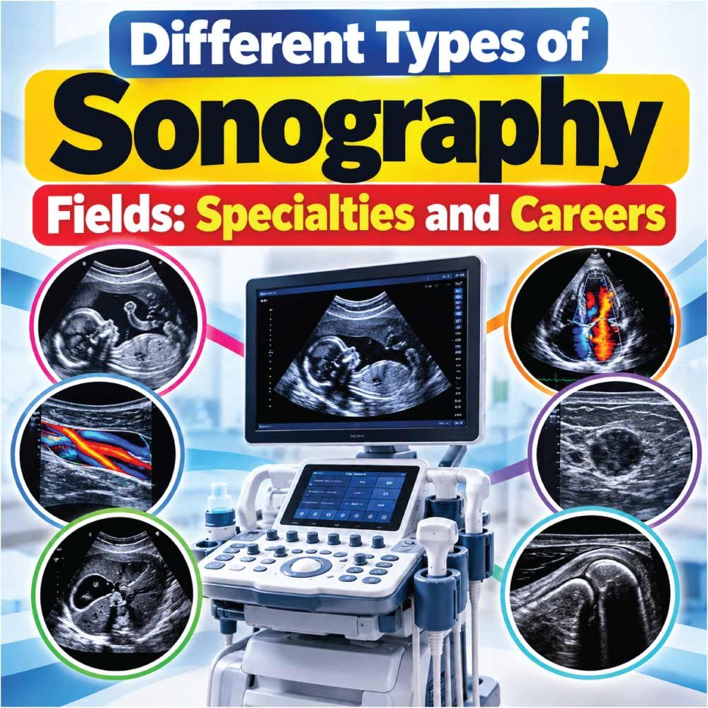
Sonography is much more than the ultrasound scans commonly associated with pregnancy. It is a diverse healthcare profession with several specialized fields, each focused on different organs, body systems, patient groups, and diagnostic needs. From examining the heart and blood vessels to imaging abdominal organs, muscles, breasts, and developing fetuses, sonographers can build expertise in many areas of medical imaging.
The different types of sonography fields also offer very different day-to-day experiences. Some specialties involve fast-paced hospital environments, while others are primarily based in outpatient clinics or specialized imaging departments. The patients, scanning techniques, equipment, anatomy, and clinical knowledge required can vary considerably from one specialty to another.
For students considering a career in diagnostic medical sonography, understanding these specialties is an important first step. This guide explores the major sonography fields, what each specialty involves, the skills required, typical work settings, and factors to consider when choosing a path that matches your interests and career goals.
What Are the Different Types of Sonography Fields?
Sonography is a broad healthcare profession that uses high-frequency sound waves to create images of structures inside the body. Although many people associate sonographers primarily with pregnancy ultrasounds, diagnostic medical sonography covers far more than obstetric imaging.
Sonographers can specialize in areas such as the abdomen, heart, blood vessels, breast, musculoskeletal system, or developing fetus. Each specialty involves different anatomy, scanning techniques, patient populations, and clinical questions.
Choosing among the different types of sonography fields can therefore make a significant difference in a sonographer’s daily work. Some specialties involve emergency patients and fast-paced hospital environments, while others may involve scheduled outpatient examinations and more predictable workflows.
Common sonography specialties include:
- Abdominal sonography
- Obstetric and gynecologic sonography
- Cardiac or echocardiography
- Vascular sonography
- Breast sonography
- Musculoskeletal sonography
- Pediatric sonography
- Neurosonography
- Other specialized and emerging ultrasound applications
Understanding these fields can help students, healthcare professionals, and people considering a sonography career identify the type of work that best matches their interests and abilities.
Abdominal Sonography
Abdominal sonography is one of the most established areas of diagnostic medical ultrasound. It focuses on organs and structures within the abdomen and can help healthcare providers investigate pain, abnormal laboratory findings, suspected disease, or other clinical concerns.
During an abdominal ultrasound, a sonographer may examine structures such as the liver, gallbladder, pancreas, spleen, kidneys, and abdominal aorta. Depending on the examination, additional surrounding structures may also be assessed.
The work requires a strong understanding of anatomy because the sonographer must obtain appropriate views and document relevant findings for interpretation by a physician or other qualified provider.
What Does an Abdominal Sonographer Do?
A typical examination may involve:
- Reviewing the patient’s history and examination request
- Explaining the procedure
- Positioning the patient appropriately
- Selecting suitable transducers and imaging settings
- Obtaining images in multiple planes
- Taking measurements when required
- Documenting the examination
- Sending images and relevant information for professional interpretation
Abdominal ultrasound can be particularly interesting for people who enjoy anatomy and diagnostic problem-solving.
Obstetric and Gynecologic Sonography
Obstetric and gynecologic sonography, often called OB/GYN sonography, focuses on the female reproductive system and pregnancy-related imaging.
Obstetric examinations may be performed at different stages of pregnancy. Depending on the clinical indication and stage of pregnancy, ultrasound can be used to evaluate fetal development, fetal anatomy, growth, location, amniotic fluid, placenta, and other aspects of pregnancy.
Gynecologic ultrasound can involve evaluation of structures such as the uterus, endometrium, ovaries, and adnexal regions.
What Makes OB/GYN Sonography Different?
This specialty combines technical imaging skills with patient communication. Pregnancy can be an emotional experience, and patients may have questions or concerns during an examination.
A sonographer’s role is to perform the examination according to professional standards and the requested protocol while maintaining appropriate communication and patient care. Diagnostic interpretation and communication of medical results remain the responsibility of the appropriate healthcare provider.
People interested in OB/GYN sonography often enjoy working with patients while developing a detailed understanding of pelvic anatomy and fetal development.
Cardiac Sonography and Echocardiography
Cardiac sonography is another major ultrasound specialty. Professionals working in this area are commonly known as cardiac sonographers or echocardiographers.
Instead of concentrating primarily on abdominal or pelvic anatomy, cardiac sonographers use ultrasound to examine the heart and its function.
Echocardiography can provide information about cardiac structures, chamber dimensions, valves, blood flow, and aspects of heart function. Doppler techniques are especially important because they allow sonographers to assess the movement of blood through the cardiovascular system.
What Does a Cardiac Sonographer Examine?
Depending on the examination, cardiac imaging may assess:
- Heart chambers
- Heart valves
- Cardiac muscle
- Blood flow
- Major cardiac vessels
- Wall motion
- Other structural and functional characteristics
Cardiac sonography requires an understanding of cardiovascular anatomy and physiology as well as specialized imaging techniques.
It can be a good fit for someone who enjoys physiology, technology, and detailed diagnostic examinations.
Vascular Sonography
Vascular sonography focuses on blood vessels rather than primarily imaging internal organs. It commonly uses grayscale ultrasound and Doppler technology to evaluate blood flow and vascular structures.
A vascular sonographer may examine arteries and veins in different parts of the body. Examinations can be performed for a variety of clinical reasons, including evaluation of suspected circulation problems.
How Does Vascular Ultrasound Work?
Vascular studies often combine structural imaging with Doppler information. The sonographer may evaluate the appearance of a vessel and then use Doppler techniques to assess characteristics of blood movement.
Common areas of vascular imaging include:
- Carotid arteries
- Upper-extremity vessels
- Lower-extremity arteries
- Lower-extremity veins
- Abdominal vessels
- Other peripheral vascular structures
Vascular sonography requires careful technique because Doppler measurements can be affected by factors such as the angle of insonation, patient positioning, and technical settings.
People interested in vascular imaging often enjoy physiology, anatomy, and examinations that involve quantitative measurements.
Breast Sonography
Breast ultrasound is a specialized application of diagnostic ultrasound used to evaluate breast tissue. It may be performed as part of a diagnostic examination or used alongside other forms of breast imaging.
Ultrasound can help characterize breast findings and distinguish different tissue appearances. It is particularly useful because it can provide real-time imaging without ionizing radiation.
Breast sonography requires careful scanning technique and attention to detail. The sonographer may document the location and characteristics of an area of interest and obtain images according to the examination protocol.
Who May Be Interested in Breast Sonography?
This field may appeal to professionals who prefer detailed examinations and patient-focused outpatient imaging.
Because breast examinations can cause anxiety for some patients, communication and professionalism are important parts of the role. Sonographers must explain the examination clearly while remaining within their professional scope.
Musculoskeletal Sonography
Musculoskeletal, or MSK, sonography uses ultrasound to examine muscles, tendons, ligaments, joints, and other superficial soft-tissue structures.
This specialty has applications in evaluating sports injuries, soft-tissue problems, inflammation, fluid collections, and other conditions where ultrasound may provide useful dynamic information.
One distinctive feature of musculoskeletal ultrasound is that structures can often be examined while the body part is moving.
Why Is Dynamic Imaging Important?
Some musculoskeletal structures behave differently during movement than when the patient is completely still. Dynamic ultrasound can therefore provide information that may complement a static examination.
An MSK sonographer needs a detailed understanding of:
- Musculoskeletal anatomy
- Tendon and muscle structure
- Joint anatomy
- Probe positioning
- Dynamic scanning
- Ultrasound artifacts
- Image optimization
MSK sonography can be particularly attractive to people interested in anatomy, sports medicine, orthopedics, and rehabilitation-related imaging.
Pediatric Sonography
Pediatric sonography involves ultrasound examinations performed on infants, children, and adolescents. Because pediatric patients differ considerably from adults, this field requires more than simply applying adult scanning techniques to younger patients.
Children may have difficulty remaining still or may feel uncomfortable around unfamiliar equipment. Sonographers therefore need patience, communication skills, and the ability to adapt their approach.
Pediatric ultrasound can involve multiple body systems, including the abdomen, pelvis, urinary system, brain in appropriate infants, and other areas depending on the clinical indication.
Skills Needed in Pediatric Ultrasound
Successful pediatric sonographers often need to be comfortable with:
- Age-specific anatomy
- Patient positioning
- Communication with children
- Communication with parents or guardians
- Appropriate transducer selection
- Maintaining attention during examinations
- Working carefully with smaller anatomical structures
The variety of examinations can make pediatric sonography a broad specialty rather than a single narrowly defined scanning area.
Neurosonography
Neurosonography is a specialized area of ultrasound involving the nervous system. In certain clinical situations, ultrasound can be used to examine structures of the brain, particularly in infants whose skull anatomy allows suitable acoustic access.
Cranial ultrasound is commonly associated with neonatal and infant imaging. It can be useful in evaluating certain intracranial structures and abnormalities in appropriate clinical settings.
Because the available acoustic windows change as the skull develops, this specialty requires knowledge of developmental anatomy and specialized scanning techniques.
Neurosonography is therefore more specialized than general abdominal or pelvic ultrasound and may be pursued by professionals working in settings where neonatal or pediatric imaging is routinely performed.
Also Read:
Specialized Sonography Fields at a Glance
The major specialties differ in anatomy, patients, techniques, and work environment. The following comparison provides a practical overview.
| Sonography Field | Main Focus | Common Work Settings | Key Skills |
|---|---|---|---|
| Abdominal | Abdominal organs and structures | Hospitals, imaging centers | Anatomy, scanning technique |
| OB/GYN | Pregnancy and reproductive anatomy | Hospitals, clinics, imaging centers | Pelvic anatomy, patient communication |
| Cardiac | Heart and cardiac function | Hospitals, cardiology departments | Cardiac anatomy, Doppler |
| Vascular | Arteries, veins, blood flow | Hospitals, vascular labs | Doppler, vascular physiology |
| Breast | Breast tissue | Breast centers, hospitals, clinics | Detailed scanning, documentation |
| Musculoskeletal | Muscles, tendons, joints | Clinics, specialty practices | Dynamic imaging, anatomy |
| Pediatric | Imaging in children | Children’s hospitals, hospitals | Pediatric anatomy, communication |
| Neurosonography | Specialized nervous-system imaging | Neonatal and pediatric settings | Developmental anatomy, specialized technique |
The boundaries between fields are not always absolute. A sonographer may work in more than one specialty, depending on education, credentials, employer requirements, and local scope of practice.
How Sonography Specialties Differ From One Another
Although every sonography specialty uses ultrasound technology, the actual work can feel very different from one field to another.
Differences in Anatomy
The first major difference is the anatomy being examined.
An abdominal sonographer needs extensive knowledge of visceral organs. A cardiac sonographer focuses heavily on the heart and cardiovascular physiology, while a vascular sonographer studies blood vessels and hemodynamics.
Musculoskeletal sonography, on the other hand, requires detailed knowledge of tendons, muscles, ligaments, and joints.
Differences in Patient Populations
Patient populations also vary.
Some specialties involve patients of all ages, while others concentrate on specific groups. Pediatric sonography focuses on younger patients, whereas OB/GYN sonography frequently involves women receiving reproductive or pregnancy-related imaging.
Cardiac and vascular departments may see patients with a wide range of cardiovascular concerns.
Differences in Work Environment
The workplace can also influence the experience of a specialty.
Sonographers may work in:
- Hospitals
- Outpatient imaging centers
- Physician offices
- Specialty clinics
- Cardiology departments
- Vascular laboratories
- Women’s health centers
- Pediatric hospitals
- Mobile imaging services
The same specialty can therefore have different working conditions depending on the employer and patient population.
Also Read:
Which Sonography Specialty Is Right for You?
There is no single specialty that is suitable for every aspiring sonographer. The right choice depends on interests, education, preferred patients, work environment, and long-term professional goals.
Instead of choosing a specialty solely because it sounds interesting, consider what you would actually enjoy doing during a typical working day.
Consider Your Interest in Anatomy
If you enjoy learning about internal organs, abdominal sonography may be appealing.
If cardiovascular physiology interests you, cardiac or vascular sonography may provide a better match.
If you enjoy movement and musculoskeletal anatomy, MSK ultrasound could be worth exploring.
Consider Your Preferred Patient Population
Think about whether you prefer:
- Adults
- Children
- Pregnant patients
- Patients with cardiovascular concerns
- Outpatient populations
- Hospital patients
- A mixture of different patient groups
Patient interaction is an important part of sonography, so this consideration should not be overlooked.
Consider the Technical Side
Different specialties place different demands on technical knowledge.
Doppler-heavy fields such as vascular and cardiac sonography require an understanding of blood flow and specialized measurements. Other areas may emphasize detailed anatomical scanning and image documentation.
Your interest in physiology, technology, anatomy, and hands-on imaging can help guide your exploration.
Also Read:
Education and Training for Sonography Specialties
Becoming a sonographer generally involves formal education and clinical training. The exact pathway depends on the specialty, employer, credentialing requirements, and country where you intend to work.
In the United States, students commonly enter accredited diagnostic medical sonography or cardiovascular technology programs. Training may include classroom instruction, laboratory practice, and supervised clinical experience.
Typical subjects can include:
- Human anatomy
- Physiology
- Ultrasound physics
- Patient care
- Medical terminology
- Pathophysiology
- Sonographic instrumentation
- Scanning techniques
- Clinical practicum
Specialization can occur through focused education, clinical experience, professional credentials, or employer-specific training.
For example, cardiac sonography follows a different educational emphasis from general diagnostic medical sonography because cardiac imaging requires specialized knowledge of cardiovascular anatomy and function.
Sonography Credentials and Specialization
Professional credentials can also differ according to the area of practice.
In the United States, organizations such as the American Registry for Diagnostic Medical Sonography (ARDMS) offer credentials associated with different areas of ultrasound. Other credentialing organizations serve specific cardiovascular or vascular professions.
Requirements can include education, clinical experience, examinations, and continuing professional development, depending on the credential.
Because credentialing rules can change and vary by jurisdiction, students should check the current requirements of the credentialing organization and employers in the area where they plan to work.
A useful approach is to identify your preferred specialty first and then investigate the education and credential pathway associated with that field.
Skills That Matter Across Sonography Fields
Despite their differences, most sonography specialties require a similar foundation of professional skills.
Technical Skills
Sonographers need to operate ultrasound equipment, select appropriate transducers, optimize images, recognize artifacts, and acquire clinically useful views.
Technical ability develops through education and repeated supervised practice.
Anatomical Knowledge
Strong anatomy knowledge is essential. A sonographer needs to understand what structures should look like and where they are located so that images can be obtained systematically.
Communication
Sonographers interact directly with patients. They need to explain examinations, obtain relevant information, provide appropriate instructions, and maintain a professional environment.
Attention to Detail
Small differences in anatomy, positioning, measurements, and image quality can matter. Careful documentation and consistent scanning techniques are therefore important in every specialty.
Physical and Professional Skills
Sonography can involve prolonged standing, repetitive movements, patient positioning, and use of the ultrasound transducer for extended periods.
Good ergonomics, safe scanning practices, teamwork, and professional communication are important parts of a sustainable career.
Can a Sonographer Work in More Than One Field?
Yes, depending on training, credentials, employer requirements, and local regulations, some sonographers work across multiple ultrasound specialties.
For example, a professional may receive training in general sonography and later develop additional expertise in vascular imaging. Another sonographer may focus primarily on OB/GYN examinations while developing experience with related imaging procedures.
Cross-training can broaden a professional’s skill set, but it requires appropriate education and clinical competence. Sonographers should not assume that experience in one specialty automatically qualifies them to perform every type of examination.
How to Explore Different Sonography Fields Before Choosing
Students do not always need to make a final specialty decision immediately. Learning about each field and observing professionals can provide a much clearer picture.
Consider taking these steps:
- Review educational programs to see which specialties are included.
- Talk with practicing sonographers about their daily responsibilities.
- Ask about clinical rotations and available specialty experiences.
- Compare work environments, such as hospitals and outpatient centers.
- Learn about credential requirements for specialties that interest you.
- Think about patient interaction and the population you prefer.
- Consider long-term career development rather than focusing only on the first job.
Clinical exposure is especially valuable because the reality of a specialty may be different from what you expect from reading about it.
The Future of Sonography Specialties
Ultrasound continues to develop as imaging technology improves and clinicians find new applications for point-of-care and specialized ultrasound.
Portable systems, improved image processing, Doppler technology, three-dimensional imaging, and expanding point-of-care ultrasound applications are influencing how ultrasound is used in healthcare.
These developments do not eliminate the importance of professional training. In fact, as ultrasound becomes available in more clinical environments, understanding anatomy, physics, limitations, image quality, and appropriate examination techniques remains essential.
Future sonographers may therefore encounter an increasingly diverse range of applications rather than a narrower set of traditional examinations.
Final Thoughts on the Different Types of Sonography Fields
The different types of sonography fields offer a wide range of professional paths. Abdominal, OB/GYN, cardiac, vascular, breast, musculoskeletal, pediatric, and neurosonography each require a distinct combination of anatomical knowledge, technical skill, patient care, and clinical experience.
The best way to explore these options is to look beyond job titles and understand what the actual work involves. Consider the anatomy you enjoy, the patients you want to work with, the technical skills you find interesting, and the clinical environment where you see yourself working.
Sonography is not simply one profession with one type of ultrasound examination. It is a collection of specialties that allows trained professionals to develop expertise in different areas of medical imaging. Understanding those differences can make education and career planning much more informed.

Zak is a dedicated medical and career writer specializing in sonography, healthcare education, and professional development. Through SonographerSalary.com, he shares in-depth insights on sonographer salaries, education pathways, and career tips to help readers build successful futures in medical imaging. His content combines accuracy with practical, easy-to-understand guidance, empowering students and professionals to make confident, informed career decisions.