
Medical imaging continues to evolve, providing healthcare professionals with better tools to diagnose and monitor diseases. One of the most significant advancements in ultrasound technology is Contrast Enhanced Ultrasound (CEUS). This specialized imaging technique improves the visibility of blood flow and tissue vascularity, helping doctors evaluate organs, tumors, and various medical conditions with greater accuracy.
Unlike traditional ultrasound, Contrast Enhanced Ultrasound uses special contrast agents that enhance the ultrasound signal, allowing clinicians to observe blood circulation in real time. Because it does not involve ionizing radiation and is generally well tolerated, CEUS has become an increasingly valuable diagnostic tool in modern medicine.
What Is Contrast Enhanced Ultrasound?
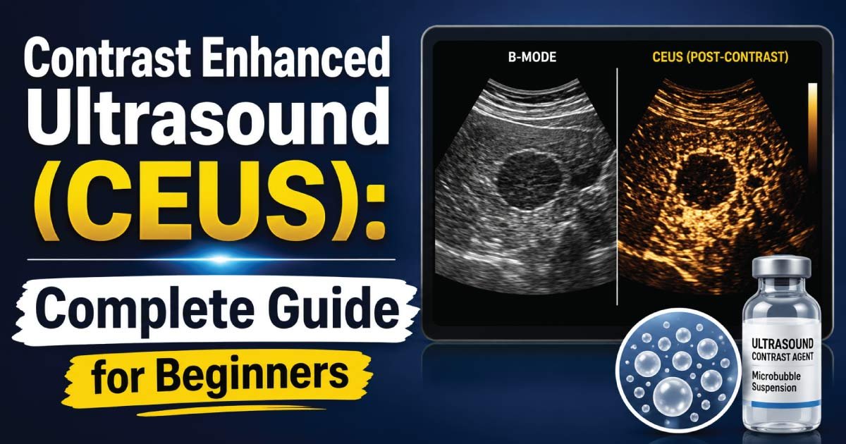
Contrast Enhanced Ultrasound (CEUS) is an advanced ultrasound imaging technique that uses tiny gas-filled microbubble contrast agents injected into a patient’s bloodstream. These microbubbles reflect ultrasound waves much more effectively than surrounding tissues and blood, creating enhanced images of blood vessels and organ perfusion.
The primary goal of CEUS is to improve visualization of blood flow and tissue vascularity that may not be clearly visible on conventional ultrasound examinations.
Healthcare providers commonly use CEUS to evaluate:
- Liver lesions
- Kidney abnormalities
- Pancreatic diseases
- Cardiac conditions
- Tumors
- Vascular disorders
- Organ transplantation monitoring
The technique allows physicians to assess how blood moves through tissues in real time, providing valuable diagnostic information.
How Contrast Enhanced Ultrasound Works
The procedure relies on specially designed microbubble contrast agents. These microscopic bubbles are usually filled with inert gases and surrounded by a protective shell.
When injected into a vein:
- The microbubbles travel through the bloodstream.
- Ultrasound waves strike the bubbles.
- The bubbles produce strong echoes.
- The ultrasound machine detects these echoes.
- Enhanced images of blood vessels and tissues are created.
Because the microbubbles remain within blood vessels rather than entering surrounding tissues, they provide excellent visualization of vascular structures and blood perfusion.
This characteristic makes CEUS particularly useful for identifying abnormal blood flow patterns associated with diseases and tumors.
What Are Ultrasound Contrast Agents?
Ultrasound contrast agents are specially formulated substances containing microscopic gas bubbles.
Characteristics include:
- Extremely small size
- Safe circulation through blood vessels
- Strong reflection of ultrasound waves
- Rapid elimination from the body
- Minimal impact on kidney function
Unlike contrast materials used in CT or MRI scans, ultrasound contrast agents are generally not toxic to the kidneys and are considered safe for many patients who cannot undergo other imaging procedures.
The body naturally eliminates the gas through the lungs after the examination.
Why Contrast Enhanced Ultrasound Is Performed
Doctors may recommend CEUS when conventional ultrasound images do not provide enough information.
Common reasons include:
Characterization of Liver Lesions
CEUS is widely used to determine whether liver lesions are benign or malignant.
It helps identify:
- Hemangiomas
- Focal nodular hyperplasia
- Hepatocellular carcinoma
- Liver metastases
The enhancement pattern of a lesion often provides important clues about its nature.
Evaluation of Tumors
Tumors often develop abnormal blood vessels.
CEUS can:
- Assess tumor vascularity
- Monitor treatment response
- Detect recurrent disease
- Guide biopsies
Assessment of Organ Perfusion
Doctors use CEUS to evaluate blood supply in organs such as:
- Liver
- Kidneys
- Pancreas
- Spleen
Poor perfusion may indicate injury, blockage, or disease.
Vascular Imaging
CEUS helps visualize:
- Blood vessels
- Aneurysms
- Vascular malformations
- Endoleaks after aneurysm repair
Transplant Monitoring
After organ transplantation, CEUS may help evaluate blood flow and detect complications early.
Advantages of Contrast Enhanced Ultrasound
CEUS offers several important benefits compared with other imaging techniques.
No Ionizing Radiation
Unlike CT scans, CEUS does not expose patients to radiation.
This makes it safer for:
- Children
- Pregnant patients in selected situations
- Individuals requiring repeated imaging
Real-Time Imaging
CEUS allows doctors to observe blood flow as it occurs.
This real-time assessment provides valuable diagnostic information that static imaging may miss.
Excellent Safety Profile
Most patients tolerate ultrasound contrast agents very well.
Serious adverse reactions are rare.
Kidney-Friendly Alternative
Many CT and MRI contrast agents may pose risks for patients with kidney disease.
CEUS contrast agents are generally safer because they are not primarily eliminated through the kidneys.
Cost-Effective
Compared with CT and MRI, CEUS may reduce healthcare costs while providing highly useful diagnostic information.
Bedside Availability
Portable ultrasound systems allow CEUS to be performed in hospitals, intensive care units, and emergency settings.
Limitations of Contrast Enhanced Ultrasound
Although CEUS provides many advantages, it also has limitations.
Operator Dependency
Image quality depends heavily on the skill and experience of the sonographer and interpreting physician.
Limited Acoustic Windows
Excessive bowel gas, obesity, or difficult anatomy may reduce image quality.
Restricted Field of View
Unlike CT and MRI, ultrasound can only image specific regions at a time.
Short Imaging Window
Microbubble contrast agents remain effective for a limited period, requiring efficient image acquisition.
How a Contrast Enhanced Ultrasound Examination Is Performed
The procedure is similar to a routine ultrasound but includes administration of a contrast agent.
Step 1: Patient Preparation
Preparation depends on the organ being examined.
Patients may be instructed to:
- Fast for several hours
- Avoid certain foods
- Follow specific medical instructions
Step 2: Initial Ultrasound Scan
The sonographer performs a standard ultrasound examination before administering contrast.
Baseline images are recorded.
Step 3: Contrast Injection
A small intravenous line is placed.
The microbubble contrast agent is injected into a vein, usually in the arm.
Step 4: Dynamic Imaging
Immediately after injection, continuous ultrasound imaging begins.
The radiologist observes:
- Arterial phase enhancement
- Portal venous phase enhancement
- Late phase enhancement
These phases provide important diagnostic information.
Step 5: Image Analysis
The physician reviews enhancement patterns and interprets the findings.
A report is then prepared for the referring healthcare provider.
Contrast Enhanced Ultrasound of the Liver
The liver is one of the most common organs evaluated using CEUS.
Liver lesions often display characteristic enhancement patterns.
Examples include:
Benign Lesions
Benign lesions frequently demonstrate predictable enhancement characteristics.
Examples:
- Hemangioma
- Focal nodular hyperplasia
- Regenerative nodules
Malignant Lesions
Cancerous lesions often show:
- Rapid arterial enhancement
- Washout during later phases
- Irregular vascular patterns
These features help differentiate malignant tumors from benign findings.
Contrast Enhanced Ultrasound of the Kidneys
CEUS plays an important role in renal imaging.
Applications include:
- Characterizing kidney masses
- Evaluating cystic lesions
- Assessing blood flow
- Detecting infarction
- Monitoring transplanted kidneys
Because ultrasound contrast agents are not harmful to kidney function, CEUS is particularly valuable for patients with renal impairment.
Contrast Enhanced Ultrasound in Oncology
Cancer diagnosis and treatment monitoring increasingly rely on advanced imaging.
CEUS helps oncologists by:
- Detecting tumor blood supply
- Assessing treatment effectiveness
- Identifying residual disease
- Evaluating recurrence
Changes in tumor vascularity often occur before changes in tumor size, allowing earlier assessment of treatment response.
Safety and Risks of Contrast Enhanced Ultrasound
CEUS is generally considered very safe.
Most patients experience no side effects.
Possible mild reactions include:
- Headache
- Nausea
- Warm sensation
- Injection site discomfort
Serious allergic reactions are extremely uncommon.
Healthcare providers screen patients carefully before administering contrast agents.
Emergency equipment is available in the rare event of a severe reaction.
CEUS vs Conventional Ultrasound
| Feature | Conventional Ultrasound | Contrast Enhanced Ultrasound |
|---|---|---|
| Blood Flow Visualization | Limited | Excellent |
| Tissue Perfusion Assessment | Limited | Detailed |
| Tumor Characterization | Moderate | Superior |
| Contrast Agent Required | No | Yes |
| Radiation Exposure | None | None |
| Real-Time Imaging | Yes | Yes |
CEUS significantly improves the diagnostic capabilities of standard ultrasound examinations.
Future of Contrast Enhanced Ultrasound
The future of CEUS is promising.
Researchers continue exploring new applications in:
- Cancer imaging
- Cardiology
- Neurology
- Organ transplantation
- Drug delivery systems
Advances in ultrasound technology and contrast agents may further improve diagnostic accuracy and expand clinical applications.
Artificial intelligence and machine learning may also help physicians analyze CEUS images more efficiently in the future.
Conclusion
Contrast Enhanced Ultrasound (CEUS) is a powerful imaging technique that enhances traditional ultrasound by using microbubble contrast agents to visualize blood flow and tissue perfusion in real time. It provides valuable diagnostic information for evaluating liver lesions, kidney diseases, tumors, vascular abnormalities, and organ transplants.
The procedure offers numerous advantages, including the absence of radiation exposure, excellent safety, real-time imaging capabilities, and suitability for patients with kidney impairment. As medical imaging technology continues to advance, Contrast Enhanced Ultrasound is expected to play an increasingly important role in modern diagnostic medicine, helping healthcare providers make more accurate and timely clinical decisions.
👉 Discover the Doppler Effect and learn how this important ultrasound principle helps measure blood flow, detect vascular conditions, and improve diagnostic accuracy. This complete guide explains Doppler ultrasound, blood flow imaging, and key clinical applications in simple terms. Read the full post to gain a deeper understanding of Doppler Effect in ultrasound and its real-world medical uses.

Zak is a dedicated medical and career writer specializing in sonography, healthcare education, and professional development. Through SonographerSalary.com, he shares in-depth insights on sonographer salaries, education pathways, and career tips to help readers build successful futures in medical imaging. His content combines accuracy with practical, easy-to-understand guidance, empowering students and professionals to make confident, informed career decisions.