
The FAST Exam is one of the most important emergency ultrasound procedures used in trauma medicine. FAST stands for Focused Assessment with Sonography for Trauma. It is a rapid bedside ultrasound examination designed to identify internal bleeding and other life-threatening injuries in trauma patients.
Emergency physicians, trauma surgeons, and sonographers use the FAST exam to quickly assess patients who have suffered accidents, falls, blunt trauma, or penetrating injuries. Because it is fast, non-invasive, and can be performed at the patient’s bedside, the FAST exam has become a standard component of modern trauma care.
This examination helps healthcare providers make critical treatment decisions within minutes, often improving patient outcomes and reducing delays in life-saving interventions.
What Is a FAST Exam?
A FAST Exam is a focused ultrasound evaluation performed primarily on trauma patients to detect free fluid within body cavities. In trauma settings, the presence of free fluid is often assumed to be blood until proven otherwise.
The exam is designed to answer a simple but vital question:
Is there internal bleeding that requires immediate treatment?
Unlike a complete abdominal ultrasound, the FAST exam focuses only on specific areas where blood commonly accumulates after traumatic injuries.
The examination can usually be completed in less than five minutes, making it an invaluable diagnostic tool in emergency situations.
Meaning of FAST
FAST stands for:
Focused Assessment with Sonography for Trauma
Each part of the name reflects the purpose of the examination:
- Focused – Targets specific body regions.
- Assessment – Evaluates for signs of injury.
- Sonography – Uses ultrasound imaging.
- Trauma – Intended for injured patients.
The FAST exam is not meant to replace comprehensive imaging studies but serves as a rapid screening tool during emergency evaluation.
Why Is the FAST Exam Performed?
The FAST exam is performed when healthcare providers suspect internal injuries following trauma.
Common situations include:
- Motor vehicle accidents
- Motorcycle accidents
- Falls from height
- Sports injuries
- Assaults
- Penetrating trauma
- Industrial accidents
- Crush injuries
The primary goal is to identify internal bleeding quickly so that treatment can begin without delay.
History of the FAST Exam
Before ultrasound became widely available in emergency departments, doctors often relied on invasive procedures or exploratory surgery to detect internal bleeding.
The introduction of FAST ultrasound revolutionized trauma care because it allowed clinicians to visualize potential bleeding sites rapidly and safely.
Over time, improvements in ultrasound technology increased the accuracy and reliability of the examination.
Today, FAST is considered a standard component of trauma assessment worldwide.
How the FAST Exam Works
Ultrasound uses high-frequency sound waves to create images of internal organs and body structures.
During the FAST exam:
- A transducer sends sound waves into the body.
- Sound waves reflect off tissues and organs.
- Returning echoes are processed by the ultrasound machine.
- Real-time images appear on the monitor.
Blood and other fluids appear differently from surrounding tissues, allowing clinicians to identify abnormal fluid collections.
The entire examination is performed at the bedside, often while other trauma care measures are simultaneously underway.
Areas Examined During a FAST Exam
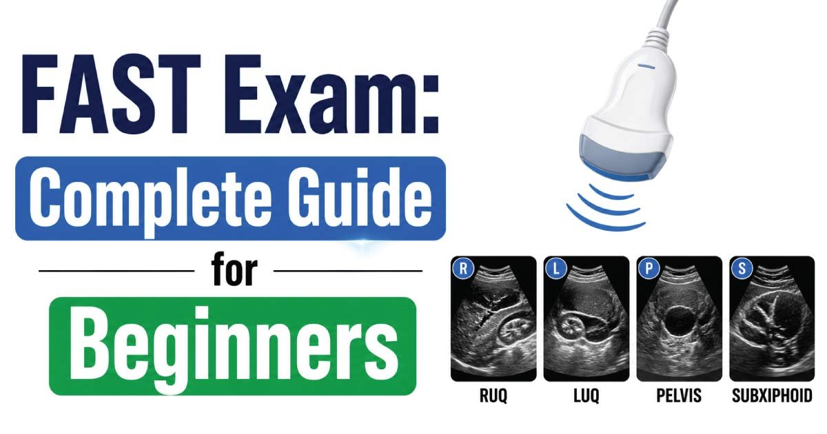
The FAST exam focuses on four primary anatomical regions where free fluid commonly collects.
Right Upper Quadrant (RUQ)
This view evaluates the space between:
- Liver
- Right kidney
This area is called Morison’s Pouch.
Because gravity causes fluid to accumulate here, it is one of the most sensitive locations for detecting internal bleeding.
A positive finding appears as a dark fluid collection between the liver and kidney.
Left Upper Quadrant (LUQ)
The left upper quadrant view examines the space between:
- Spleen
- Left kidney
This region may contain blood following splenic injury or other abdominal trauma.
The sonographer carefully evaluates this area for abnormal fluid accumulation.
Pelvic View
The pelvic examination assesses the lowest part of the abdominal cavity.
Fluid often collects here due to gravity.
In males, fluid may accumulate behind the bladder.
In females, fluid may collect within the rectouterine pouch.
This view is particularly useful when only a small amount of bleeding is present.
Subxiphoid Cardiac View
The cardiac window evaluates the heart and surrounding pericardial sac.
This portion of the exam helps identify:
- Pericardial effusion
- Cardiac tamponade
- Traumatic heart injuries
Cardiac tamponade is a life-threatening condition in which fluid compresses the heart and interferes with normal function.
Early detection can be lifesaving.
What Is an Extended FAST Exam (eFAST)?
Modern trauma centers frequently perform an Extended FAST (eFAST) examination.
The eFAST expands the traditional FAST exam by evaluating the lungs and pleural spaces.
Additional assessments include:
- Pneumothorax
- Hemothorax
- Pleural effusions
This expanded approach allows clinicians to identify chest injuries in addition to abdominal trauma.
FAST Exam Views Explained
The examination consists of several standardized ultrasound windows.
Morison’s Pouch View
This view evaluates the hepatorenal recess.
It is often the first location where free intraperitoneal blood becomes visible.
Splenorenal View
This image assesses the relationship between the spleen and left kidney.
Blood appears as an anechoic or dark collection.
Pelvic View
Both transverse and longitudinal views are typically obtained.
The sonographer examines areas surrounding the bladder for fluid accumulation.
Cardiac View
The subxiphoid approach provides a clear view of the heart.
This view helps identify pericardial fluid and assess cardiac motion.
Indications for FAST Exam
Healthcare providers perform FAST examinations for many clinical situations.
Common indications include:
- Blunt abdominal trauma
- Penetrating trauma
- Unexplained shock
- Hypotension
- Multiple injuries
- Suspected internal bleeding
- Cardiac injury
- Chest trauma
The exam is especially useful in unstable patients who cannot safely undergo lengthy imaging procedures.
Advantages of the FAST Exam
FAST has become popular because of its many benefits.
Rapid Assessment
The examination can be completed within minutes.
This speed is crucial during trauma emergencies.
Non-Invasive
No surgical procedures or needles are required for imaging.
No Radiation Exposure
Unlike CT scans, FAST does not expose patients to ionizing radiation.
Portable
Ultrasound machines can be used virtually anywhere.
Examples include:
- Emergency departments
- Intensive care units
- Ambulances
- Helicopter transport
- Battlefield settings
Repeatable
The exam can be repeated multiple times without risk.
This allows clinicians to monitor changes over time.
Cost-Effective
FAST is generally less expensive than advanced imaging studies.
Limitations of the FAST Exam
Despite its usefulness, FAST has limitations.
Operator Dependency
Accuracy depends heavily on the experience of the examiner.
Limited Detection of Organ Injuries
The exam primarily detects fluid rather than directly visualizing organ damage.
Small Bleeds May Be Missed
Very small amounts of blood may not be detectable during early injury stages.
Obesity and Bowel Gas
Excess body fat and intestinal gas can reduce image quality.
Cannot Replace CT Scans
CT imaging remains superior for detailed evaluation of many traumatic injuries.
FAST is best viewed as a rapid screening tool.
What Is a Positive FAST Exam?
A positive FAST exam indicates the presence of abnormal free fluid.
In trauma patients, this fluid is generally considered blood until proven otherwise.
Positive findings may appear as:
- Dark fluid collections
- Fluid surrounding organs
- Pericardial fluid
- Pleural fluid
A positive examination often prompts immediate surgical consultation or further imaging.
What Is a Negative FAST Exam?
A negative FAST exam means no obvious free fluid is detected.
However, a negative study does not completely rule out injury.
Some injuries:
- Bleed slowly
- Produce minimal fluid
- Occur outside examined regions
Patients may require repeat FAST examinations or CT imaging if symptoms persist.
Role of FAST Exam in Emergency Medicine
The FAST exam plays a central role in trauma management.
It assists clinicians in:
- Rapid triage
- Surgical decision-making
- Monitoring unstable patients
- Identifying life-threatening injuries
- Guiding emergency interventions
Its ability to provide immediate information makes it one of the most valuable ultrasound applications in emergency medicine.
Training Required for FAST Examination
Healthcare professionals performing FAST examinations typically undergo specialized training.
Common providers include:
- Emergency physicians
- Trauma surgeons
- Critical care physicians
- Sonographers
- Military medical personnel
Training includes image acquisition, anatomy recognition, and interpretation of abnormal findings.
Regular practice improves accuracy and confidence.
Future of FAST Ultrasound
Advances in ultrasound technology continue to improve FAST examinations.
Emerging developments include:
- Artificial intelligence assistance
- Portable handheld ultrasound devices
- Improved image quality
- Tele-ultrasound consultation
- Enhanced trauma protocols
These innovations may further increase the speed and effectiveness of trauma assessment worldwide.
Conclusion
The FAST Exam (Focused Assessment with Sonography for Trauma) is a rapid, non-invasive ultrasound examination used to identify internal bleeding and life-threatening injuries in trauma patients. By evaluating key anatomical regions such as Morison’s pouch, the splenorenal recess, pelvis, and heart, clinicians can quickly determine whether emergency intervention is required.
Its speed, portability, safety, and effectiveness have made FAST a cornerstone of modern trauma care. Although it does not replace CT scanning, it remains one of the most valuable bedside diagnostic tools in emergency and critical care medicine. As ultrasound technology continues to advance, the FAST exam will likely play an even greater role in saving lives and improving trauma outcomes.
👉 Learn everything about Aliasing in Ultrasound with this comprehensive guide. Understand what causes aliasing, how it affects Doppler ultrasound imaging, and the best techniques to reduce or prevent it. Whether you’re a student or healthcare professional, this guide explains key concepts in simple language. Read the full post to master ultrasound aliasing and improve your diagnostic knowledge.

Zak is a dedicated medical and career writer specializing in sonography, healthcare education, and professional development. Through SonographerSalary.com, he shares in-depth insights on sonographer salaries, education pathways, and career tips to help readers build successful futures in medical imaging. His content combines accuracy with practical, easy-to-understand guidance, empowering students and professionals to make confident, informed career decisions.