
The Doppler effect in ultrasound is the change in frequency of sound waves when they reflect off moving objects, such as blood cells. It helps measure direction and speed of blood flow in vessels. This principle is widely used in Doppler ultrasound to assess circulation and detect vascular abnormalities accurately.
The Doppler Effect is one of the most important principles in physics and medical ultrasound. It explains how the frequency of sound waves changes when there is relative motion between a sound source and an observer. This phenomenon is widely used in everyday life, weather forecasting, astronomy, radar systems, and especially in diagnostic medical imaging.
In healthcare, the Doppler Effect forms the foundation of Doppler ultrasound, allowing doctors and sonographers to evaluate blood flow, heart function, and circulation throughout the body. Understanding the Doppler Effect can help patients, students, and healthcare professionals better understand how modern ultrasound technology works.
In this complete guide, we will explain what the Doppler Effect is, how it works, its history, applications in medicine, and why it is so important in diagnostic imaging.
What Is the Doppler Effect?
The Doppler Effect is the change in frequency or wavelength of a wave as the source of the wave and the observer move relative to each other.
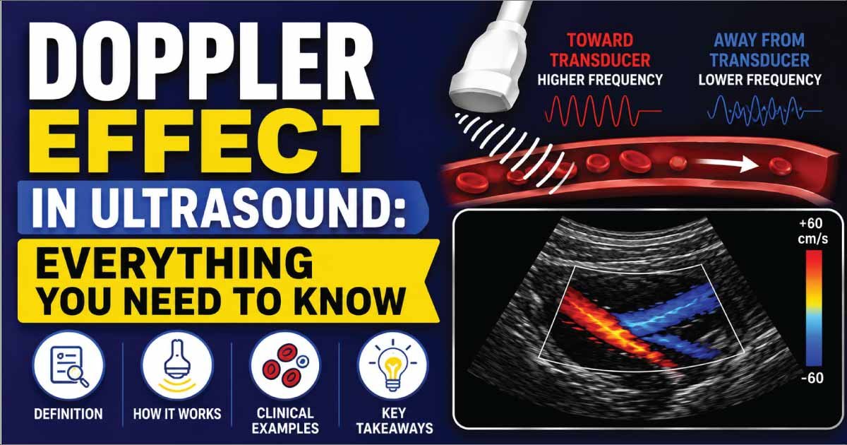
When an object producing waves moves toward an observer, the waves become compressed, causing the observed frequency to increase. When the object moves away, the waves spread out, causing the observed frequency to decrease.
In simple terms, the Doppler Effect causes waves to appear higher or lower in frequency depending on movement.
This phenomenon occurs with:
- Sound waves
- Light waves
- Radio waves
- Ultrasound waves
Who Discovered the Doppler Effect?
The Doppler Effect was first described in 1842 by:
Christian Doppler
Christian Doppler theorized that the frequency of waves changes when the source and observer move relative to one another.
His discovery eventually became essential for:
- Medical ultrasound
- Astronomy
- Weather radar
- Satellite communication
- Speed detection systems
Today, the Doppler Effect remains one of the most widely applied concepts in wave physics.
A Simple Example of the Doppler Effect
A common example is an ambulance passing by on a road.
As the Ambulance Approaches
- Sound waves compress.
- Frequency increases.
- The siren sounds higher in pitch.
As the Ambulance Passes and Moves Away
- Sound waves spread apart.
- Frequency decreases.
- The siren sounds lower in pitch.
Although the actual sound emitted by the siren never changes, the observed frequency changes because of relative motion.
This is the Doppler Effect in action.
How Does the Doppler Effect Work?
Waves travel through a medium at a specific speed.
When the source is stationary:
- Wavefronts are evenly spaced.
- The observer receives a constant frequency.
When the source moves:
- Wavefronts compress in front of the source.
- Wavefronts stretch behind the source.
As a result:
- Higher frequency is detected in front.
- Lower frequency is detected behind.
This frequency change is known as the Doppler Shift.
Doppler Effect Formula
The Doppler Effect can be expressed mathematically.
For medical ultrasound, the Doppler frequency shift is represented by:
f_D=\frac{2f_0v\cos\theta}{c}
Where:
- fD = Doppler frequency shift
- f0 = Transmitted ultrasound frequency
- v = Velocity of blood flow
- θ = Doppler angle
- c = Speed of sound in tissue
This formula allows ultrasound systems to calculate blood flow velocity.
What Is Doppler Shift?
Doppler Shift refers specifically to the difference between the transmitted frequency and the received frequency.
It is the measurable result of the Doppler Effect.
In ultrasound:
- Blood moving toward the transducer creates a positive shift.
- Blood moving away creates a negative shift.
The ultrasound machine analyzes this shift to determine blood flow characteristics.
Positive Doppler Effect
A positive Doppler Effect occurs when the source or reflecting object moves toward the observer.
Characteristics include:
- Increased frequency
- Compressed wavefronts
- Higher observed pitch
In medical ultrasound, blood moving toward the transducer produces a positive Doppler shift.
Negative Doppler Effect
A negative Doppler Effect occurs when the source or reflecting object moves away from the observer.
Characteristics include:
- Reduced frequency
- Expanded wavefronts
- Lower observed pitch
In Doppler ultrasound, blood flowing away from the transducer creates a negative Doppler shift.
The Doppler Effect in Medical Ultrasound
Medical ultrasound uses the Doppler Effect to analyze blood flow.
The ultrasound transducer:
- Sends sound waves into the body.
- Receives echoes reflected from moving red blood cells.
- Measures frequency changes.
- Calculates blood velocity and direction.
This allows doctors to evaluate circulation throughout the body.
Why Is the Doppler Effect Important in Medicine?
The Doppler Effect provides information that conventional ultrasound cannot.
It helps healthcare providers:
- Measure blood flow speed
- Determine blood flow direction
- Evaluate heart function
- Detect vascular disease
- Monitor pregnancies
- Assess organ circulation
Without the Doppler Effect, ultrasound would only provide structural images.
Types of Doppler Ultrasound
Several ultrasound techniques rely on the Doppler Effect.
Color Doppler
Color Doppler displays blood flow using color maps.
Common applications include:
- Artery evaluation
- Vein assessment
- Pregnancy imaging
- Cardiac studies
Spectral Doppler
Spectral Doppler displays blood flow velocity as waveforms.
It is useful for:
- Measuring blood velocity
- Detecting stenosis
- Assessing vascular disease
Power Doppler
Power Doppler detects weak or slow blood flow.
It is often used when conventional color Doppler is insufficient.
Pulsed Wave Doppler
Pulsed Wave Doppler measures blood flow at a specific location.
It is widely used in vascular imaging.
Continuous Wave Doppler
Continuous Wave Doppler measures very high blood velocities.
It is frequently used in echocardiography.
Doppler Effect in Cardiology
Cardiac ultrasound relies heavily on Doppler technology.
Doctors use Doppler studies to evaluate:
- Heart valves
- Blood flow through chambers
- Valve stenosis
- Valve regurgitation
- Congenital heart defects
Doppler findings are critical for diagnosing many heart conditions.
Doppler Effect in Vascular Ultrasound
Vascular sonographers use the Doppler Effect to examine:
- Carotid arteries
- Peripheral arteries
- Deep veins
- Renal arteries
- Abdominal vessels
These studies help identify:
- Blood clots
- Vessel narrowing
- Reduced circulation
- Vascular abnormalities
Doppler Effect During Pregnancy
Obstetric Doppler ultrasound evaluates blood flow between the mother, placenta, and baby.
Common assessments include:
- Umbilical artery flow
- Placental circulation
- Fetal blood supply
- Fetal heart function
These examinations help ensure healthy fetal development.
Also Read:
Doppler Effect in Neurology
The Doppler Effect is also used to evaluate blood flow within the brain.
Examples include:
- Transcranial Doppler studies
- Cerebral circulation monitoring
- Stroke assessment
These applications help doctors evaluate neurological conditions.
Doppler Effect in Weather Forecasting
Outside medicine, the Doppler Effect is widely used in meteorology.
Weather radar systems analyze Doppler shifts to detect:
- Storm movement
- Wind speed
- Tornado formation
- Rainfall patterns
This information improves weather prediction accuracy.
Doppler Effect in Astronomy
Astronomers use the Doppler Effect to study stars and galaxies.
It helps determine:
- Whether objects are moving toward Earth
- Whether objects are moving away from Earth
- Galaxy motion
- Expansion of the universe
One of the most important discoveries in modern astronomy relies on Doppler principles.
Factors Affecting the Doppler Effect
The Doppler effect in ultrasound is influenced by several physical and technical factors that determine the accuracy of blood flow measurements. These variables affect how frequency shifts are produced and interpreted, making correct technique essential for reliable diagnostic results.
1. Velocity of Motion
The speed of moving structures, especially blood cells, directly affects the Doppler shift. Faster blood flow produces larger frequency changes, while slower flow results in smaller shifts, helping clinicians estimate the intensity of circulation within vessels.
2. Wave Frequency
The frequency of the transmitted ultrasound wave plays a major role in Doppler sensitivity. Higher transmitted frequencies generate larger measurable shifts, improving detection of slow or small-volume blood flow in superficial vessels and fine vascular structures.
3. Direction of Movement
The direction of blood flow relative to the ultrasound probe determines the nature of the Doppler shift. Movement toward the transducer produces a positive shift, while movement away results in a negative shift, helping define flow orientation.
4. Doppler Angle
The Doppler angle is the angle between the ultrasound beam and the direction of blood flow. Accurate measurements require an optimal angle (usually less than 60 degrees), as incorrect alignment can lead to significant errors in velocity calculation.
Also Read:
Advantages of Doppler Technology
Doppler ultrasound technology provides significant advantages in medical imaging by allowing real-time evaluation of blood flow without invasive procedures. It enhances diagnostic accuracy while remaining safe, accessible, and cost-effective for routine and advanced clinical use.
1. Non-Invasive
Doppler ultrasound is completely non-invasive, meaning no surgical procedures or internal instruments are required. This makes it a safe and comfortable diagnostic tool for patients while still providing detailed information about blood circulation and vascular health.
2. Safe
Doppler imaging does not use ionizing radiation, unlike CT scans or X-rays. This makes it safe for repeated use in patients of all ages, including pregnant women, without exposing them to harmful radiation effects.
3. Real-Time Results
One of the major advantages of Doppler technology is its ability to provide instant, real-time information about blood flow. Clinicians can immediately assess vascular conditions, detect abnormalities, and make quick clinical decisions during the examination.
4. Cost-Effective
Ultrasound and Doppler studies are generally more affordable compared to advanced imaging techniques such as MRI or CT scans. This makes them widely used in both developed and resource-limited healthcare settings for routine diagnostic evaluation.
5. Widely Available
Doppler ultrasound machines are available in most hospitals and diagnostic centers. Its accessibility allows physicians to quickly perform vascular assessments, making it a standard and essential tool in everyday clinical practice across many medical specialties.
Limitations of Doppler Ultrasound
Doppler ultrasound is a valuable diagnostic tool, but it also has important limitations that can affect accuracy and image interpretation. These limitations are mainly related to technical factors, operator skill, and the physical behavior of ultrasound waves in different tissues.
1. Angle Dependency
Doppler measurements are highly dependent on the angle between the ultrasound beam and blood flow. Incorrect probe positioning can lead to inaccurate velocity readings, making proper alignment essential for reliable and precise results.
2. Operator Dependence
The accuracy of Doppler ultrasound strongly depends on the skill and experience of the operator. Poor technique, incorrect settings, or improper probe handling can lead to misinterpretation of blood flow patterns and diagnostic errors.
3. Limited Through Bone and Air
Ultrasound waves do not travel well through dense bone or air-filled structures. This limitation reduces Doppler effectiveness in certain body regions, such as areas behind bone or gas-filled organs, restricting full vascular assessment.
4. Potential Artifacts
Doppler imaging can be affected by artifacts such as aliasing, noise, or motion interference. These artifacts may distort true blood flow patterns and create challenges in accurate interpretation, especially in complex or low-flow vascular conditions.
Common Doppler-Related Terms
You may encounter these related terms:
- Doppler Effect
- Doppler Shift
- Color Doppler
- Power Doppler
- Spectral Doppler
- Aliasing
- Blood Flow Velocity
- Resistive Index
- Pulsed Wave Doppler
- Continuous Wave Doppler
Understanding these terms can make ultrasound reports easier to interpret.
Also Read:
Clinical Significance of the Doppler Effect
The Doppler Effect transformed ultrasound from a purely anatomical imaging tool into a functional diagnostic system capable of measuring blood flow.
Today, it plays a vital role in:
- Cardiology
- Vascular medicine
- Obstetrics
- Neurology
- Emergency medicine
Healthcare providers use Doppler information every day to diagnose disease, monitor treatment, and improve patient outcomes.
Conclusion
The Doppler Effect is the change in wave frequency that occurs when there is relative motion between a wave source and an observer. This principle forms the foundation of Doppler ultrasound and enables healthcare professionals to measure blood flow velocity, direction, and circulation throughout the body.
From evaluating heart valves and blood vessels to monitoring fetal health during pregnancy, the Doppler Effect has become an essential component of modern medical imaging. Beyond medicine, it is also widely used in weather forecasting, astronomy, radar systems, and many other scientific fields.
Understanding the Doppler Effect helps patients, students, and healthcare professionals appreciate the science behind one of the most powerful diagnostic tools in healthcare today.

Zak is a dedicated medical and career writer specializing in sonography, healthcare education, and professional development. Through SonographerSalary.com, he shares in-depth insights on sonographer salaries, education pathways, and career tips to help readers build successful futures in medical imaging. His content combines accuracy with practical, easy-to-understand guidance, empowering students and professionals to make confident, informed career decisions.