
Aliasing in Doppler ultrasound is a display error that occurs when blood flow velocity exceeds the system’s sampling limit (Nyquist limit). It causes inaccurate wrapping of the waveform, often showing reversed or distorted flow. Adjusting scale settings or using a lower frequency can help reduce aliasing and improve diagnostic accuracy.
Ultrasound technology has transformed modern healthcare by allowing doctors and sonographers to visualize organs, tissues, and blood flow in real time. One of the most valuable applications of ultrasound is Doppler imaging, which measures the direction and speed of blood flow within the body.
However, Doppler ultrasound is not perfect. Various artifacts can affect image quality and blood flow measurements. One of the most common Doppler ultrasound artifacts is aliasing. Understanding aliasing is important because it can influence how blood flow appears on an ultrasound image and may sometimes mimic or hide medical conditions.
In this complete guide, we will explain what aliasing is, how it occurs, why it happens, how it appears on Doppler ultrasound, and the methods sonographers use to reduce or eliminate it.
What Is Aliasing?
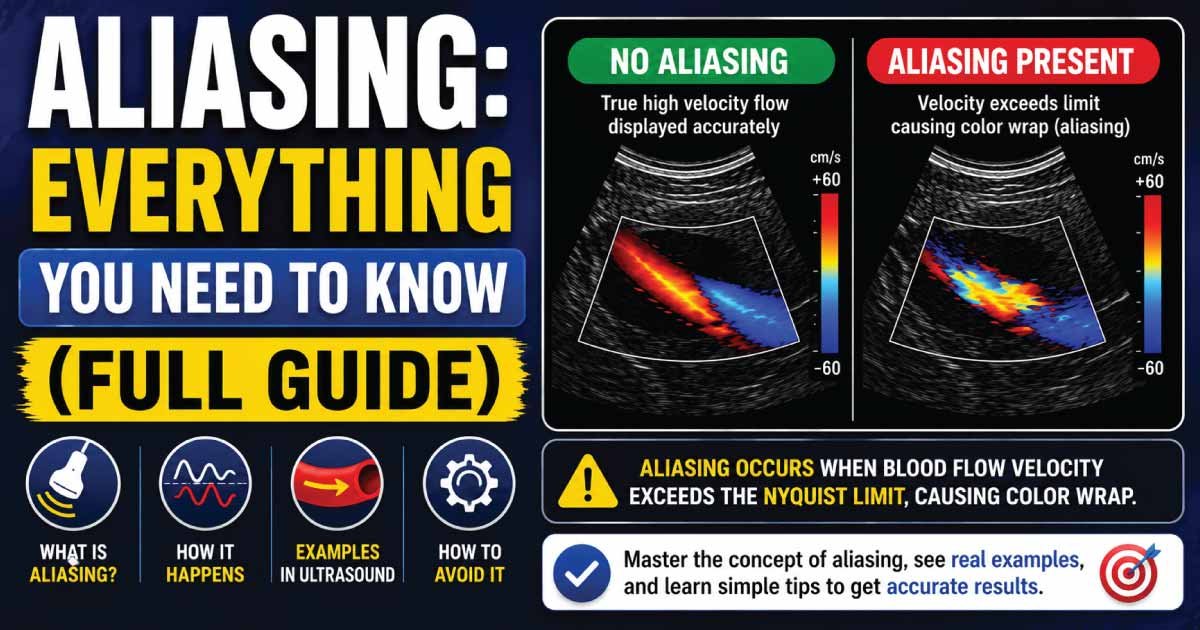
Aliasing is a Doppler ultrasound artifact that occurs when blood flow velocity exceeds the maximum velocity that the ultrasound system can accurately measure.
When this happens, the Doppler signal becomes misrepresented, causing blood flow to appear in the wrong direction or at an incorrect velocity.
In simple terms, aliasing occurs when the ultrasound machine cannot correctly process very high blood flow speeds.
As a result, the displayed Doppler information becomes distorted.
Why Does Aliasing Occur?
Aliasing occurs because of limitations in the sampling rate used by Doppler ultrasound systems.
Pulsed Wave Doppler sends ultrasound pulses into the body and listens for returning echoes between pulses.
If blood is moving too fast, the reflected frequency shift exceeds the system’s ability to measure it accurately.
The ultrasound machine then incorrectly interprets the signal, producing aliasing.
This phenomenon is closely related to the Nyquist Limit, which determines the maximum Doppler shift that can be measured without distortion.
Understanding the Nyquist Limit
The Nyquist Limit is the highest Doppler frequency shift that a pulsed Doppler system can accurately detect.
It is equal to one-half of the Pulse Repetition Frequency (PRF).
Nyquist\ Limit=\frac{PRF}{2}
When the Doppler frequency shift exceeds the Nyquist Limit:
- Aliasing occurs.
- Velocity measurements become inaccurate.
- Blood flow may appear reversed.
- Color Doppler displays become distorted.
The Nyquist Limit is one of the most important concepts in Doppler ultrasound.
How Does Aliasing Appear on Ultrasound?
The appearance of aliasing depends on the Doppler mode being used.
Color Doppler Aliasing
In Color Doppler imaging, aliasing appears as an abrupt color change.
For example:
- Red flow may suddenly turn blue.
- Blue flow may suddenly turn red.
- Mosaic color patterns may appear.
This creates a confusing display of blood flow direction.
Spectral Doppler Aliasing
In Spectral Doppler, aliasing appears when the waveform extends beyond the display limits and wraps around to the opposite side of the baseline.
Characteristics include:
- Waveform cutoff
- Baseline crossing
- Signal wraparound
- Velocity distortion
This is one of the most recognizable signs of aliasing.
How Does Aliasing Affect Doppler Measurements?
Aliasing can affect the accuracy of Doppler examinations by:
- Misrepresenting blood velocity
- Incorrectly displaying blood flow direction
- Making interpretation more difficult
- Masking true blood flow characteristics
Experienced sonographers recognize aliasing and adjust imaging settings accordingly.
Where Is Aliasing Commonly Seen?
Aliasing is a common Doppler ultrasound artifact that occurs when blood flow exceeds the system’s ability to accurately measure velocity. It is most often seen in areas of stenosis, turbulence, or abnormal cardiovascular flow patterns where blood moves at very high speeds.
1. Heart Valves
Aliasing is frequently observed across heart valves, especially when they are narrowed or diseased. Conditions such as valve stenosis cause blood to accelerate rapidly, producing high-velocity flow that exceeds Doppler limits and results in aliasing.
2. Carotid Arteries
In the carotid arteries, aliasing commonly occurs in cases of significant atherosclerotic narrowing. The reduced vessel diameter increases blood flow speed, leading to turbulent high-velocity jets that produce characteristic Doppler aliasing patterns.
3. Peripheral Arteries
Peripheral arterial disease can cause stenosis in limb arteries, resulting in increased blood velocity at narrowed segments. This high-speed flow often exceeds the Doppler scale, making aliasing a useful indicator of vascular obstruction.
4. Renal Arteries
Renal artery stenosis is another important site where aliasing may be seen. Narrowing of the renal arteries leads to elevated flow velocities, which can appear as color reversal or wrapping artifacts on Doppler imaging.
5. Congenital Heart Defects
Congenital heart abnormalities can create abnormal blood flow pathways with extremely high velocities. These altered flow dynamics frequently produce aliasing, helping clinicians identify structural and functional cardiac defects during echocardiographic evaluation.
Aliasing in Color Doppler Imaging
Color Doppler uses color coding to represent blood flow.
Typically:
- Red indicates flow toward the transducer.
- Blue indicates flow away from the transducer.
When aliasing occurs:
- Colors abruptly reverse.
- Mixed color patterns appear.
- Flow may seem turbulent.
Although this can complicate interpretation, it often alerts the examiner to the presence of very high blood flow velocities.
Aliasing in Spectral Doppler
Spectral Doppler displays blood flow velocity over time.
Under normal circumstances:
- Positive flow appears above the baseline.
- Negative flow appears below the baseline.
When aliasing occurs:
- The waveform exceeds the display limit.
- Part of the waveform wraps around.
- Velocity values become inaccurate.
This is particularly common when evaluating severe arterial stenosis.
What Causes Aliasing?
Aliasing occurs in Doppler ultrasound when blood flow velocity exceeds the system’s ability to accurately sample and display frequency shifts. This leads to a misleading reversal or wrapping of the waveform, commonly seen in high-velocity or stenotic blood flow conditions.
1. High Blood Flow Velocity
High blood flow velocity is the most common cause of aliasing. When blood moves too fast, the Doppler shift exceeds the Nyquist limit, causing incorrect display of flow direction or waveform distortion on the ultrasound screen.
2. Low Pulse Repetition Frequency (PRF)
A low PRF reduces the Nyquist limit, making it easier for aliasing to occur. Since PRF controls sampling rate, lower values cannot accurately capture high-velocity blood flow, leading to signal overlap and incorrect representation.
3. High Ultrasound Frequency
Higher transducer frequencies generate larger Doppler shifts for the same blood flow velocity. This increases the likelihood of exceeding the system’s detection limit, especially in vessels with already elevated flow speeds.
4. Deep Vessel Location
Deep vessels often require lower PRF settings to improve signal penetration. However, this adjustment reduces the measurable velocity range, increasing the risk of aliasing in deeper anatomical structures.
5. Improper Doppler Settings
Incorrect machine settings such as inappropriate gain, scale, or baseline positioning can contribute to aliasing. Poor optimization of Doppler parameters may distort true flow patterns and reduce diagnostic accuracy.
Also Read:
How Can Aliasing Be Reduced?
Sonographers use several strategies to minimize or eliminate aliasing and improve image accuracy. These adjustments help increase the measurable velocity range and improve visualization of blood flow patterns.
1. Increase Pulse Repetition Frequency (PRF)
Increasing PRF raises the Nyquist limit, allowing the system to measure higher blood flow velocities without distortion. This is one of the most effective ways to reduce aliasing.
2. Shift the Baseline
Adjusting the Doppler baseline creates more display space for high-velocity flow in one direction. This helps reduce waveform wrapping and improves visualization of directional blood flow.
3. Use a Lower Frequency Transducer
Lower-frequency transducers produce smaller Doppler shifts, which reduces the chance of exceeding the Nyquist limit. This improves accuracy in cases of high-velocity flow.
4. Reduce Doppler Angle
Reducing the angle between the ultrasound beam and blood flow decreases the measured frequency shift. This helps bring velocities within the detectable range and minimizes aliasing effects.
5. Use Continuous Wave Doppler
Continuous Wave Doppler is not limited by the Nyquist constraint and therefore does not produce aliasing. It is especially useful in evaluating very high-velocity flows, such as in severe stenosis or valvular disease.
Continuous Wave Doppler and Aliasing
Continuous Wave Doppler differs from Pulsed Wave Doppler.
Because it continuously transmits and receives ultrasound waves:
- There is no Nyquist Limit restriction.
- Very high velocities can be measured.
- Aliasing does not occur.
However, Continuous Wave Doppler cannot precisely identify the depth of the measured flow.
Clinical Importance of Aliasing
Although aliasing is technically an artifact, it often provides useful clinical information.
Its presence may indicate:
- Severe vessel narrowing
- High blood flow velocity
- Valve disease
- Turbulent flow
- Congenital abnormalities
Radiologists and sonographers frequently use aliasing as a clue to identify abnormal blood flow.
Aliasing in Cardiac Ultrasound
Echocardiography commonly demonstrates aliasing.
Examples include:
- Aortic stenosis
- Mitral regurgitation
- Tricuspid regurgitation
- Pulmonary stenosis
Aliasing often helps identify areas of abnormal cardiac blood flow.
Also Read:
Aliasing in Vascular Ultrasound
Vascular sonographers routinely encounter aliasing when evaluating:
- Carotid artery stenosis
- Peripheral arterial disease
- Renal artery stenosis
- Arteriovenous fistulas
The presence of aliasing often suggests increased blood velocity.
Aliasing During Pregnancy Ultrasound
Obstetric Doppler studies may occasionally show aliasing in:
- Umbilical arteries
- Fetal vessels
- Placental circulation
These findings are carefully evaluated to ensure normal fetal blood flow.
Advantages of Recognizing Aliasing
Recognizing aliasing in Doppler ultrasound is clinically important because it provides valuable clues about underlying blood flow conditions. When correctly identified, it improves diagnostic accuracy and helps guide further vascular assessment.
1. Identify High-Velocity Flow
Aliasing often indicates increased blood flow velocity. This can be a sign of physiological acceleration or pathological conditions such as stenosis, where blood moves faster through narrowed vessels.
2. Improve Diagnostic Accuracy
Correct recognition of aliasing prevents misinterpretation of Doppler waveforms. Understanding this artifact helps clinicians distinguish true flow abnormalities from technical display limitations, improving overall diagnostic confidence.
3. Detect Vascular Disease
Aliasing is frequently associated with vascular narrowing or obstruction. Its presence can help identify conditions like arterial stenosis or turbulent flow, prompting further investigation and clinical evaluation.
4. Guide Further Testing
When aliasing is observed, additional Doppler adjustments or advanced imaging may be required. This helps clinicians obtain more accurate velocity measurements and a clearer understanding of vascular health.
Limitations of Aliasing
Although aliasing provides useful diagnostic information, it also has several limitations that can affect interpretation and accuracy in Doppler ultrasound studies.
1. Distorts Velocity Measurements
Aliasing can cause blood flow velocity to appear incorrectly displayed. This distortion makes it difficult to determine the true speed of blood flow, especially in high-velocity vessels.
2. Complicates Interpretation
For less experienced operators, aliasing can be confusing and may be mistaken for abnormal flow patterns. This increases the risk of misinterpretation and requires proper training for accurate diagnosis.
3. Requires Technical Adjustments
To correct aliasing, ultrasound machine settings such as PRF, baseline, or frequency often need adjustment. This makes the examination more technically demanding and dependent on operator expertise.
4. Can Mask True Flow Patterns
In severe cases, aliasing may completely obscure the actual blood flow pattern. This can limit the ability to fully assess vascular conditions and may require additional imaging techniques for clarification.
Also Read:
Common Doppler Terms Related to Aliasing
When studying Doppler ultrasound, you may encounter:
- Doppler Effect
- Doppler Shift
- Nyquist Limit
- Pulse Repetition Frequency (PRF)
- Color Doppler
- Spectral Doppler
- Continuous Wave Doppler
- Pulsed Wave Doppler
- Blood Flow Velocity
- Turbulent Flow
Understanding these concepts helps explain why aliasing occurs.
When Should You Be Concerned About Aliasing?
Patients generally do not need to worry about aliasing itself.
Aliasing is not a disease.
Instead, it is an ultrasound artifact that may indicate:
- Fast blood flow
- Vessel narrowing
- Cardiac abnormalities
- Other circulatory conditions
Your healthcare provider will interpret the finding in the context of your overall examination.
Conclusion
Aliasing is a common Doppler ultrasound artifact that occurs when blood flow velocity exceeds the maximum measurable limit of a Pulsed Wave Doppler system. It results from the Doppler frequency shift surpassing the Nyquist Limit, causing blood flow to be displayed incorrectly.
Although aliasing can distort Doppler measurements, it is often a valuable diagnostic clue that helps healthcare professionals identify high-velocity blood flow, vascular narrowing, and cardiac abnormalities. By understanding aliasing and the factors that cause it, sonographers can adjust imaging settings and obtain more accurate diagnostic information.
As one of the most frequently encountered Doppler artifacts, aliasing remains an important concept for students, sonographers, and healthcare professionals involved in ultrasound imaging.

Zak is a dedicated medical and career writer specializing in sonography, healthcare education, and professional development. Through SonographerSalary.com, he shares in-depth insights on sonographer salaries, education pathways, and career tips to help readers build successful futures in medical imaging. His content combines accuracy with practical, easy-to-understand guidance, empowering students and professionals to make confident, informed career decisions.