
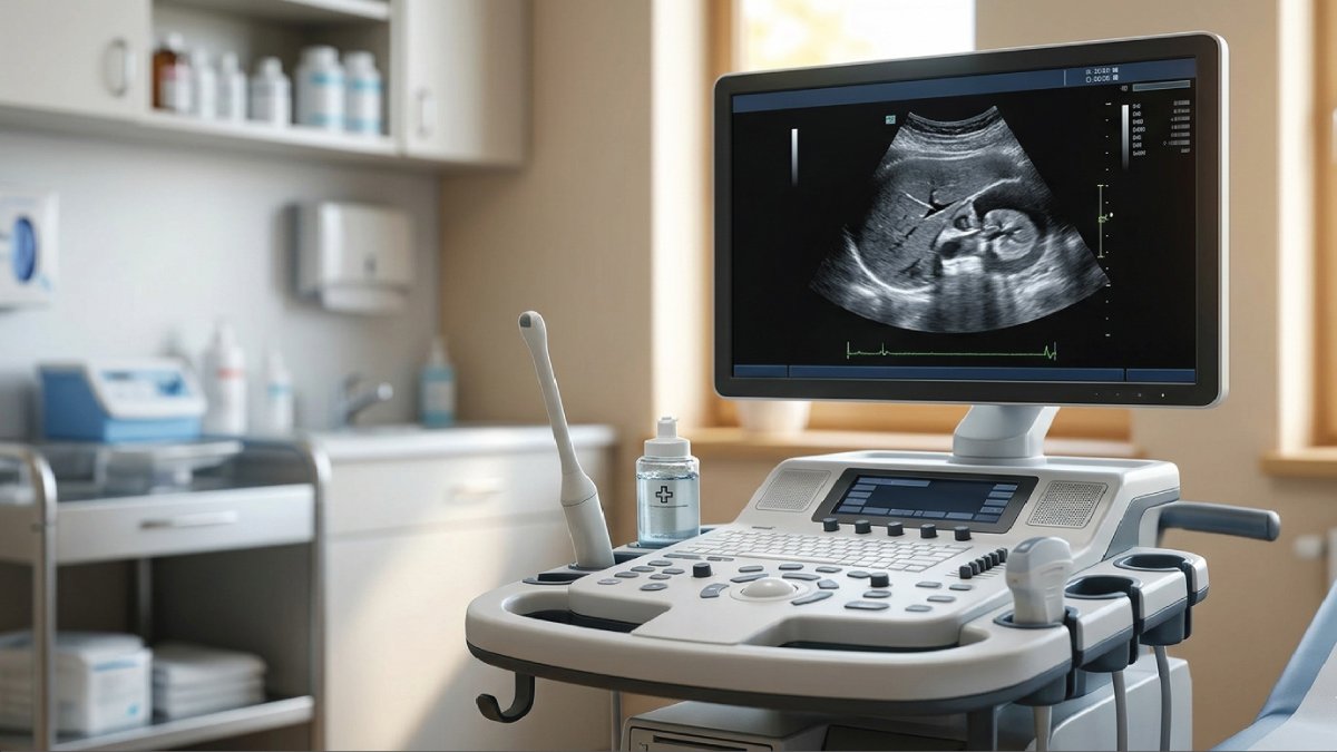
Diagnostic medical sonography is one of the most important and rapidly growing fields in modern healthcare. If you have ever seen an ultrasound image of a baby during pregnancy or heard about imaging tests that do not involve radiation, you have already encountered this technology.
But beyond those familiar examples, there is much more to understand about this fascinating profession and medical tool.In this comprehensive guide, we will break down what is diagnostic medical sonography, how it works, why it matters, and what career opportunities it offers all while naturally exploring key concepts like diagnostic sonography meaning, medical sonography definition, and ultrasound diagnostic field.
What Is Diagnostic Medical Sonography?
At its core, diagnostic medical sonography is a medical imaging technique that uses high-frequency sound waves to produce images of structures inside the body. These images help doctors diagnose and monitor a wide range of medical conditions.
So, if you are asking what is diagnostic medical ultrasound, it is essentially the same thing just another term used interchangeably with diagnostic medical sonography. Both refer to the use of ultrasound technology for medical imaging purposes.
Medical Sonography Definition
A simple medical sonography definition would be:
A non-invasive imaging method that uses sound waves to create visual images of internal organs, tissues, and blood flow.
Unlike X-rays or CT scans, sonography does not use ionizing radiation, making it a safer option for many patients, including pregnant women.
Diagnostic Sonography Meaning Explained
To fully understand the diagnostic sonography meaning, it is helpful to break the term into its basic parts and see how they work together in medical practice. This helps learners and patients clearly understand what the process actually involves in healthcare settings today.
- Diagnostic → Used to identify or monitor medical conditions
- Sonography → Imaging using sound waves
Put together, diagnostic sonography refers to using sound-based imaging to assist in medical diagnosis.
This sonography explanation highlights why it’s such a valuable tool it provides real-time imaging that helps healthcare providers make accurate and timely decisions.
How Does Diagnostic Medical Sonography Work?
Understanding how diagnostic medical sonography works helps make the technology behind imaging clearer and easier to follow. It is based on the use of high-frequency sound waves that travel through the body and return as echoes, which are then processed into detailed visual images used for medical evaluation.
The Basic Process
- A device called a transducer is placed on the patient’s skin
- It emits high-frequency sound waves into the body
- These waves bounce off internal structures
- The returning echoes are captured and converted into images
These images are displayed on a monitor in real time, allowing medical professionals to observe movement, blood flow, and organ function.
Why Sound Waves?
Sound waves are used because they offer a safe and efficient way to see inside the body without radiation or invasive procedures.
- Are safe and non-invasive
- Provide real-time imaging
- Can penetrate soft tissues effectively
This makes sonography ideal for examining organs like the heart, liver, kidneys, and uterus.
Types of Diagnostic Medical Sonography
The ultrasound diagnostic field is highly diverse, offering several specialized branches that focus on different parts of the body and specific clinical needs. Each type of sonography plays an important role in helping healthcare professionals diagnose conditions accurately and choose the most effective treatment approach for patients.
1. Obstetric and Gynecologic Sonography
Monitors pregnancy progression and fetal development throughout different stages
Examines female reproductive organs to detect abnormalities or health conditions
2. Abdominal Sonography
Evaluates major abdominal organs such as the liver, pancreas, kidneys, and spleen
Helps detect issues like gallstones, cysts, infections, or possible tumors
3. Cardiac Sonography (Echocardiography)
Studies the structure and overall function of the heart in real time
Helps in diagnosing heart diseases, valve problems, and blood flow irregularities
4. Vascular Sonography
Examines blood circulation within arteries and veins throughout the body
Detects blockages, narrowing of vessels, or dangerous blood clots
5. Musculoskeletal Sonography
Focuses on muscles, joints, tendons, and other soft tissue structures
Commonly used for diagnosing sports injuries and movement related conditions
These specialties highlight how broad and impactful medical imaging sonography can be.
Sonography Basics Everyone Should Know
If you are new to this topic, understanding a few core sonography basics can make the concept much easier to grasp. These fundamental points explain why ultrasound is such a widely trusted imaging method in modern healthcare and how it benefits both patients and medical professionals in everyday clinical practice.
- Non-invasive: No surgery or needles required in most cases
- Radiation-free: Safer than many imaging alternatives
- Real-time imaging: Doctors can see movement instantly
- Widely used: From emergency rooms to prenatal care
These fundamentals make sonography one of the most versatile diagnostic tools in healthcare.
Also Read:
Diagnostic Ultrasound Meaning in Modern Medicine
The diagnostic ultrasound meaning in modern medicine extends beyond simple imaging, as it represents a major advancement in safe and efficient healthcare delivery. It allows doctors to examine internal organs and monitor health conditions without invasive procedures, improving both patient safety and diagnostic accuracy.
Key Advantages
- Early detection of diseases
- Guidance for medical procedures (like biopsies)
- Monitoring ongoing conditions
- Reduced healthcare costs compared to more complex imaging
Because of these benefits, the use of diagnostic ultrasound continues to grow worldwide.
Role of a Diagnostic Medical Sonographer
A diagnostic medical sonographer is a specially trained healthcare professional responsible for performing ultrasound examinations and capturing accurate diagnostic images. They play a key role in ensuring that imaging results are clear, useful, and properly interpreted by physicians for correct diagnosis and treatment planning.
What They Do
- Operate ultrasound equipment
- Prepare patients for procedures
- Capture and analyze images
- Work closely with physicians
They play a critical role in the diagnostic imaging sonography process, ensuring accurate results and patient comfort.
Skills Required
- Strong technical knowledge
- Attention to detail
- Communication skills
- Compassion for patients
Education and Career Path
If you are interested in joining the ultrasound diagnostic field, it is important to understand the education and training required to build a strong professional foundation. The career path involves structured academic study, hands on clinical experience, and professional certification that prepares individuals for real-world medical imaging work in healthcare environments.
1. Education
- Associate or bachelor’s degree in sonography provides the core academic foundation in anatomy, physiology, and imaging technology
- Specialized training programs focus on different sonography fields such as abdominal, cardiac, or obstetric imaging
- Clinical internships offer practical, hands-on experience working with real patients under supervision
2. Certification
Certification is often required or strongly recommended to validate skills and improve employability Many professionals obtain certification to improve job prospects, professional credibility, and career advancement opportunities in the healthcare sector.
3.Career Opportunities
-
Hospitals employ sonographers for a wide range of diagnostic imaging services
-
Clinics use ultrasound specialists for routine examinations and patient monitoring
-
Diagnostic imaging centers focus specifically on advanced imaging procedures and diagnostics
-
Private practices may hire sonographers for specialized or independent medical services
-
The demand for sonographers is steadily increasing due to growing healthcare needs, making it a stable and promising career choice for the future.
The demand for sonographers is steadily increasing, making it a promising career choice.
Also Read:
Why Diagnostic Medical Sonography Matters
The importance of understanding what is diagnostic medical sonography lies in its significant real-world impact on both patient care and medical decision making. It plays a key role in modern healthcare by offering safe, fast, and reliable imaging that helps doctors diagnose and manage a wide range of health conditions effectively.
Benefits to Patients
- Safer imaging option compared to radiation-based techniques, making it suitable for all age groups
- Quick results that help reduce waiting time and support faster treatment decisions
- Comfortable procedures that are generally painless and non-invasive, improving patient experience
Benefits to Healthcare Providers
- Accurate diagnostics that support correct identification of medical conditions
- Real-time insights that allow immediate observation of internal body activity
- Cost-effective solutions that reduce the need for more expensive imaging tests
This combination makes medical imaging sonography a cornerstone of modern medical care.
Common Uses of Sonography
Diagnostic imaging sonography is widely used in everyday medical practice because it provides quick, safe, and reliable information about what is happening inside the body. It supports doctors in diagnosing a variety of conditions and monitoring patient health without the need for invasive procedures or exposure to radiation.
- Monitoring pregnancy and fetal health
- Diagnosing abdominal pain
- Detecting tumors or cysts
- Evaluating heart conditions
- Checking blood flow issues
These applications show how versatile and essential this technology is.
Also Read:
Limitations of Sonography
While sonography is an extremely valuable and widely used imaging technique in modern medicine, it is important to understand that it also has certain limitations that can affect its diagnostic accuracy in specific situations. These limitations are mainly related to the physical properties of sound waves and the dependency on human operation during imaging.
- Not effective for imaging bones or air-filled structures
- Image quality can depend on the operators skill
- May require additional tests for confirmation
Even so, it remains one of the safest and most widely used imaging methods.
Future of the Ultrasound Diagnostic Field
The ultrasound diagnostic field is rapidly advancing as modern technology continues to improve imaging quality, speed, and accessibility. These developments are transforming how healthcare professionals use sonography, making it more accurate, portable, and integrated with digital systems for better patient care and diagnosis in different medical settings.
Emerging Trends
- 3D and 4D imaging provide more detailed and realistic views of internal structures, especially useful in pregnancy and complex organ assessments
- Portable ultrasound devices are making imaging available in emergency situations, rural areas, and even bedside examinations
- AI-assisted image analysis helps improve accuracy by supporting doctors in detecting abnormalities faster and more consistently
- Tele-sonography (remote imaging) allows ultrasound scans to be shared and reviewed by specialists from different locations
These innovations are expanding the capabilities of diagnostic medical ultrasound, making it even more accessible and powerful.
Final Thoughts
So, what is diagnostic medical sonography? It is much more than just an imaging technique it is a vital tool that supports accurate diagnosis, improves patient care, and advances modern medicine.
From understanding the diagnostic sonography meaning to exploring sonography basics, this field offers both practical medical benefits and exciting career opportunities. Whether you’re a student, patient, or healthcare enthusiast, knowing the medical sonography definition and its applications helps you appreciate its role in saving lives and improving health outcomes.
As technology continues to evolve, medical imaging sonography will only become more important, shaping the future of diagnostics and patient care worldwide.

Zak is a dedicated medical and career writer specializing in sonography, healthcare education, and professional development. Through SonographerSalary.com, he shares in-depth insights on sonographer salaries, education pathways, and career tips to help readers build successful futures in medical imaging. His content combines accuracy with practical, easy-to-understand guidance, empowering students and professionals to make confident, informed career decisions.