
Time Gain Compensation (TGC) is one of the most important image optimization controls in ultrasound imaging. It allows sonographers to adjust image brightness at specific depths, ensuring that structures throughout the image appear with appropriate echogenicity and detail.
Without Time Gain Compensation, deeper tissues would often appear darker than superficial tissues because ultrasound waves lose energy as they travel through the body. TGC compensates for this loss, helping produce a uniform and diagnostic ultrasound image.
Understanding Time Gain Compensation is essential for sonography students, ultrasound professionals, and anyone learning how ultrasound equipment works. This guide explains the principles, functions, applications, advantages, and common mistakes associated with TGC.
What Is Time Gain Compensation (TGC)?
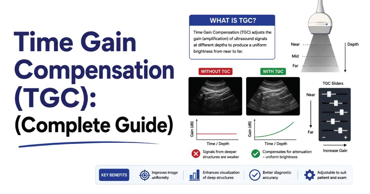
Time Gain Compensation (TGC) is an ultrasound machine control that adjusts the amplification of returning echoes based on their depth within the body.
As ultrasound waves travel deeper into tissues, they become weaker due to attenuation. The echoes returning from deep structures therefore have lower amplitudes and appear darker on the ultrasound image.
TGC selectively increases amplification for echoes returning from deeper tissues, helping create a balanced image from top to bottom.
Definition of Time Gain Compensation
Time Gain Compensation can be defined as:
A depth-specific amplification control that compensates for attenuation by increasing echo brightness at selected depths within an ultrasound image.
Its primary purpose is to maintain consistent image brightness throughout the scanning field.
Why Is Time Gain Compensation Important?
Ultrasound beams lose energy as they travel through tissue.
This process occurs because of:
- Absorption
- Reflection
- Refraction
- Scattering
As depth increases:
- Echo strength decreases
- Signal intensity weakens
- Image brightness drops
Without compensation, deeper anatomy would be difficult to visualize.
TGC helps restore visibility by amplifying these weaker returning echoes.
How Does Time Gain Compensation Work?
TGC works by increasing receiver gain according to the time required for echoes to return.
Since deeper echoes take longer to return:
- Superficial echoes receive less amplification
- Deep echoes receive greater amplification
The ultrasound machine calculates the travel time of echoes and applies varying levels of gain at different depths.
This process produces a more uniform grayscale image.
Understanding Attenuation
To understand TGC, it is important to understand attenuation.
What Is Attenuation?
Attenuation refers to the reduction of ultrasound beam intensity as it travels through tissue.
Causes include:
- Absorption
- Reflection
- Scattering
- Refraction
The deeper the ultrasound beam travels, the greater the attenuation.
Effects of Attenuation
Attenuation causes:
- Darker deep tissues
- Reduced image quality
- Loss of detail
- Poor visualization of anatomy
TGC is specifically designed to compensate for these effects.
Where Is the TGC Control Located?
Most ultrasound machines contain a vertical row of sliders known as TGC controls.
These sliders are usually located on the control panel.
Each slider corresponds to a specific depth range within the image.
The upper sliders control:
- Near-field echoes
- Superficial tissues
The lower sliders control:
- Deep echoes
- Far-field structures
By moving the sliders individually, sonographers can fine-tune image brightness at different depths.
TGC vs Overall Gain
Many beginners confuse TGC with overall gain.
Although both controls affect image brightness, they function differently.
| Feature | TGC | Overall Gain |
|---|---|---|
| Controls Specific Depths | Yes | No |
| Controls Entire Image | No | Yes |
| Adjusts Selected Areas | Yes | No |
| Improves Depth Uniformity | Yes | Limited |
| Used for Fine Tuning | Yes | No |
Overall gain changes brightness throughout the entire image, while TGC adjusts brightness at individual depths.
How to Adjust TGC Properly
Correct TGC adjustment is essential for producing diagnostic images.
Step 1: Obtain the Image
Acquire a standard ultrasound image of the target anatomy.
Step 2: Evaluate Brightness Distribution
Observe whether:
- The top is too bright
- The bottom is too dark
- Certain depths appear uneven
Step 3: Adjust Individual Sliders
Increase gain in dark areas.
Decrease gain in excessively bright areas.
Step 4: Create Uniform Brightness
Aim for a smooth transition from superficial to deep tissues.
Step 5: Reassess the Image
Ensure anatomy remains visible without excessive brightness or darkness.
Appearance of Proper TGC Adjustment
When TGC is adjusted correctly:
- Tissue brightness appears balanced
- Anatomy is clearly visualized
- No depth region appears excessively dark
- No depth region appears excessively bright
- Diagnostic information is preserved
Proper TGC contributes significantly to image quality.
Appearance of Incorrect TGC Adjustment
Excessive Deep Gain
If lower TGC sliders are increased too much:
- Deep tissues become overly bright
- Noise increases
- Artifacts may appear
Insufficient Deep Gain
If lower TGC sliders are too low:
- Deep structures appear dark
- Important anatomy may be missed
Excessive Near Gain
When upper sliders are too high:
- Superficial tissues become overexposed
- Detail is lost
Insufficient Near Gain
When upper sliders are too low:
- Near-field structures appear excessively dark
Clinical Applications of TGC
Time Gain Compensation is used in virtually every ultrasound examination.
Abdominal Ultrasound
TGC improves visualization of:
- Liver
- Gallbladder
- Pancreas
- Kidneys
- Spleen
Obstetric Ultrasound
It helps evaluate:
- Fetal anatomy
- Placenta
- Amniotic fluid
Cardiac Ultrasound
TGC improves visibility of:
- Heart chambers
- Heart valves
- Cardiac walls
Vascular Ultrasound
Proper TGC helps display:
- Vessel walls
- Blood flow
- Plaque formation
Musculoskeletal Ultrasound
It enhances imaging of:
- Tendons
- Ligaments
- Muscles
- Soft tissues
TGC in Different Tissue Types
Different tissues attenuate ultrasound differently.
Fluid-Filled Structures
Examples:
- Cysts
- Urinary bladder
- Gallbladder
These structures have minimal attenuation.
Lower TGC adjustments may be needed.
Soft Tissue
Moderate attenuation occurs.
Standard TGC settings are usually sufficient.
Dense Tissue
Examples:
- Fat
- Fibrous tissue
Greater attenuation may require increased deep gain.
Benefits of Time Gain Compensation
TGC provides several important advantages.
Improved Image Uniformity
Brightness remains consistent throughout the image.
Enhanced Diagnostic Accuracy
Structures become easier to identify.
Better Deep-Tissue Visualization
Far-field anatomy becomes more visible.
Reduced Image Artifacts
Proper adjustment minimizes certain brightness-related artifacts.
Optimized Workflow
Sonographers can quickly improve image quality.
Common TGC Patterns
Many sonographers use characteristic TGC patterns.
Straight-Line Pattern
All sliders aligned evenly.
Used when image brightness is already balanced.
Ascending Pattern
Lower sliders positioned higher.
Useful when deep tissues appear dark.
Descending Pattern
Upper sliders positioned higher.
Used less frequently.
Gentle Curve Pattern
Often considered the ideal configuration.
Provides gradual compensation with depth.
Common Mistakes When Using TGC
Using Overall Gain Instead of TGC
Many beginners increase overall gain rather than adjusting depth-specific controls.
Overcompensating Deep Tissues
Excessive deep gain creates noise and artifacts.
Ignoring Near-Field Brightness
Superficial tissues may become overexposed.
Constantly Changing TGC
Frequent unnecessary adjustments can make image interpretation difficult.
TGC and Image Quality Optimization
TGC is one of several image optimization tools.
It works alongside:
- Overall gain
- Dynamic range
- Focus position
- Frequency selection
- Harmonic imaging
Together, these controls help produce high-quality diagnostic images.
Role of Sonographers in TGC Adjustment
Sonographers are responsible for optimizing ultrasound images.
Their duties include:
- Evaluating image brightness
- Adjusting TGC settings
- Recognizing attenuation effects
- Minimizing artifacts
- Ensuring diagnostic quality
Proper TGC adjustment is considered a fundamental ultrasound scanning skill.
Future of TGC Technology
Modern ultrasound systems increasingly use automation.
Emerging developments include:
- Automatic TGC adjustment
- AI-assisted image optimization
- Adaptive gain algorithms
- Real-time brightness correction
- Smart imaging technologies
Despite automation, understanding manual TGC remains essential for sonographers.
Conclusion
Time Gain Compensation (TGC) is a critical ultrasound imaging control that compensates for attenuation by adjusting echo amplification at different depths. It helps create uniform image brightness, improves visualization of deep structures, and enhances overall diagnostic quality. Unlike overall gain, which affects the entire image, TGC allows precise depth-specific adjustments. Mastering Time Gain Compensation is an essential skill for sonographers because proper use significantly improves image quality, diagnostic confidence, and patient care across all ultrasound specialties.

Zak is a dedicated medical and career writer specializing in sonography, healthcare education, and professional development. Through SonographerSalary.com, he shares in-depth insights on sonographer salaries, education pathways, and career tips to help readers build successful futures in medical imaging. His content combines accuracy with practical, easy-to-understand guidance, empowering students and professionals to make confident, informed career decisions.