
Doppler shift refers to the change in frequency between the transmitted and received ultrasound waves when they reflect off moving blood cells. It is used in Doppler ultrasound to calculate blood flow velocity and direction. This shift helps detect vascular conditions, blockages, and abnormal circulation in medical imaging effectively.
Ultrasound technology has revolutionized modern medicine by allowing healthcare professionals to view organs, tissues, and blood flow inside the body without surgery or radiation. One of the most important principles behind Doppler ultrasound is the Doppler Shift.
The Doppler Shift helps doctors evaluate blood flow, detect vascular diseases, monitor pregnancy, assess heart function, and diagnose numerous medical conditions. Without the Doppler Shift principle, modern Doppler ultrasound would not be possible.
In this complete guide, you will learn what Doppler Shift is, how it works, why it is important, its applications in medical ultrasound, and how healthcare professionals use it to evaluate blood circulation and organ function.
What Is Doppler Shift?
Doppler Shift refers to the change in frequency of a sound wave when the source of the sound or the object reflecting the sound is moving relative to the observer.
In medical ultrasound, the ultrasound transducer sends sound waves into the body. When these waves strike moving blood cells, the reflected sound waves return with a slightly different frequency.
This difference between the transmitted frequency and the received frequency is called the Doppler Shift.
In simple terms, Doppler Shift occurs because blood cells are moving while the ultrasound machine is sending and receiving sound waves.
Who Discovered the Doppler Effect?
The Doppler Effect was first described by Austrian physicist:
Christian Doppler
Christian Doppler
In 1842, Doppler proposed that the observed frequency of waves changes when the source and observer move relative to each other.
His theory later became the foundation for:
- Medical Doppler ultrasound
- Radar systems
- Astronomy
- Weather forecasting
- Traffic speed detection
Today, Doppler’s discovery remains one of the most important concepts in wave physics.
Understanding the Doppler Effect
To understand Doppler Shift, imagine hearing a passing ambulance.
As the ambulance approaches:
- Sound waves compress.
- Frequency increases.
- The siren sounds higher in pitch.
As the ambulance moves away:
- Sound waves spread apart.
- Frequency decreases.
- The siren sounds lower in pitch.
The same principle applies to ultrasound waves interacting with moving blood cells.
How Does Doppler Shift Work in Ultrasound?
A Doppler ultrasound system works through several steps:
Step 1: Transmission of Ultrasound Waves
The transducer emits high-frequency sound waves into the body.
Step 2: Reflection from Moving Blood Cells
Red blood cells moving through blood vessels reflect these waves.
Step 3: Frequency Change
Because the blood cells are moving, the reflected frequency changes.
Step 4: Detection by the Transducer
The transducer receives the reflected waves.
Step 5: Calculation of Blood Velocity
The ultrasound machine calculates the Doppler Shift and converts it into blood flow information.
This process occurs continuously in real time.
Doppler Shift Formula
The Doppler Shift used in medical ultrasound is represented by:
f_D=\frac{2f_0v\cos\theta}{c}
Where:
- fD = Doppler frequency shift
- f0 = Transmitted ultrasound frequency
- v = Blood velocity
- θ = Doppler angle
- c = Speed of sound in tissue
This formula allows ultrasound systems to estimate blood flow velocity accurately.
Why Is Doppler Shift Important?
Doppler Shift provides information that standard ultrasound cannot.
It allows healthcare professionals to:
- Measure blood flow speed
- Determine blood flow direction
- Detect vascular blockages
- Assess heart function
- Monitor fetal circulation
- Diagnose vascular diseases
Without Doppler Shift, ultrasound could only display anatomy and structure.
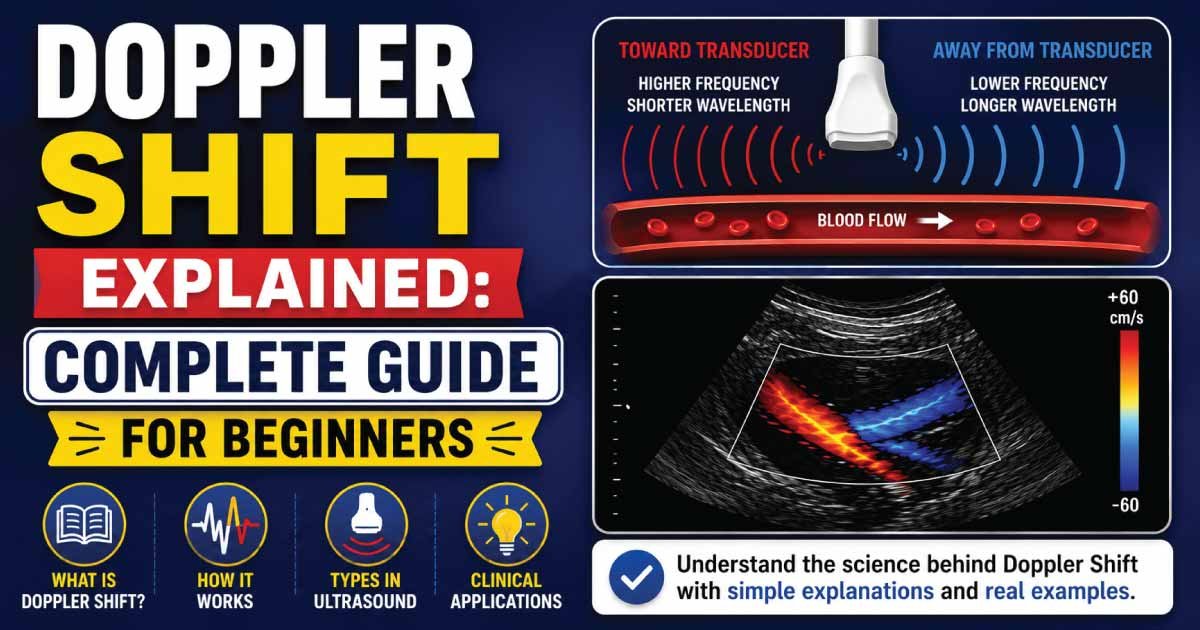
Positive Doppler Shift
A positive Doppler Shift occurs when blood flow moves toward the ultrasound transducer.
Characteristics include:
- Increased frequency
- Positive velocity values
- Blood approaching the probe
Most ultrasound systems display this flow above the baseline on spectral Doppler.
Negative Doppler Shift
A negative Doppler Shift occurs when blood moves away from the transducer.
Characteristics include:
- Decreased frequency
- Negative velocity values
- Blood moving away from the probe
This flow is often displayed below the baseline.
The Importance of Doppler Angle
Accurate Doppler measurements depend on proper angle alignment.
Ideal Doppler Angle
Most sonographers aim for:
- 30° to 60°
This range provides reliable velocity measurements.
Problems with Large Angles
As the angle approaches 90°:
- Doppler Shift decreases.
- Measurement accuracy declines.
- Velocity calculations become unreliable.
Types of Doppler Ultrasound
Several Doppler techniques use Doppler Shift principles.
Color Doppler
Displays blood flow direction and velocity using colors.
Common uses:
- Blood vessel assessment
- Pregnancy ultrasound
- Cardiac imaging
Spectral Doppler
Provides detailed velocity waveforms.
Used for:
- Vascular studies
- Cardiology
- Blood flow analysis
Power Doppler
Detects low-velocity blood flow.
Often used when standard color Doppler is insufficient.
Continuous Wave Doppler
Measures very high blood velocities.
Commonly used in echocardiography.
Pulsed Wave Doppler
Measures blood flow at specific locations.
Widely used in vascular and abdominal imaging.
Also Read:
Doppler Shift in Cardiac Ultrasound
Echocardiography relies heavily on Doppler Shift.
Doctors use it to evaluate:
- Heart valves
- Blood flow through chambers
- Valve stenosis
- Valve regurgitation
- Congenital heart defects
Doppler information is essential for many cardiac diagnoses.
Doppler Shift in Vascular Ultrasound
Vascular sonographers use Doppler Shift to examine:
- Carotid arteries
- Peripheral arteries
- Veins
- Renal arteries
- Abdominal vessels
This helps detect:
- Narrowing
- Blockages
- Blood clots
- Abnormal circulation
Doppler Shift During Pregnancy
Obstetric ultrasound frequently uses Doppler technology.
Doctors evaluate:
- Umbilical artery blood flow
- Placental circulation
- Fetal blood supply
- Fetal heart function
Doppler studies help ensure the baby receives adequate oxygen and nutrients.
Doppler Shift in Liver Imaging
Doppler ultrasound helps assess blood flow within:
- Portal vein
- Hepatic veins
- Hepatic artery
It assists in diagnosing:
- Portal hypertension
- Liver cirrhosis
- Vascular abnormalities
Doppler Shift in Kidney Ultrasound
Kidney Doppler studies evaluate:
- Renal artery flow
- Renal vein flow
- Kidney perfusion
These examinations help detect:
- Renal artery stenosis
- Kidney disease
- Vascular disorders
Advantages of Doppler Shift Technology
Doppler shift technology provides important clinical advantages by allowing non-invasive assessment of blood flow in real time. It is widely used in medical imaging because it is safe, accessible, and highly effective for diagnosing vascular conditions.
1. Non-Invasive
Doppler ultrasound is completely non-invasive, meaning no needles, incisions, or surgical procedures are required. This makes it a comfortable and safe diagnostic method for patients while still providing accurate information about blood circulation.
2. Radiation-Free
Unlike CT scans or X-rays, Doppler ultrasound does not use ionizing radiation. This makes it safe for repeated use in all patient groups, including children and pregnant women, without exposing them to harmful radiation effects.
3. Real-Time Results
Doppler technology provides immediate, real-time evaluation of blood flow. Clinicians can quickly observe vascular activity, detect abnormalities, and make on-the-spot diagnostic decisions during the ultrasound examination.
4. Cost-Effective
Ultrasound and Doppler studies are generally more affordable than advanced imaging methods such as MRI or CT. This makes them widely used in routine clinical practice and accessible in both urban and rural healthcare settings.
5. Widely Available
Doppler ultrasound machines are available in most hospitals and diagnostic centers. Their wide availability allows quick vascular assessment, making them a standard tool in emergency, outpatient, and inpatient diagnostic care.
Also Read:
Limitations of Doppler Shift
Doppler shift is a powerful diagnostic principle in ultrasound imaging, but it has several limitations that can affect accuracy and reliability. These limitations are mainly related to physical properties of sound waves, operator skill, and imaging conditions.
1. Angle Dependency
Doppler measurements are highly dependent on the angle between the ultrasound beam and blood flow. Incorrect probe positioning or poor alignment can significantly reduce accuracy and lead to underestimation or incorrect velocity readings.
2. Operator Dependence
The quality and accuracy of Doppler results strongly depend on the experience of the sonographer. Improper technique, incorrect machine settings, or poor probe handling can result in inaccurate interpretation of blood flow patterns.
3. Limited Through Bone and Air
Ultrasound waves do not pass easily through bone or air-filled structures. This limits Doppler use in certain anatomical regions and reduces the ability to evaluate blood flow in deeper or obscured areas of the body.
4. Artifact Susceptibility
Doppler imaging is prone to artifacts such as aliasing, noise, and motion-related distortions. These artifacts can interfere with accurate measurement of blood flow and may lead to misinterpretation if not properly recognized and corrected.
Common Doppler-Related Terms
When reading ultrasound reports, you may encounter:
- Doppler Shift
- Doppler Effect
- Color Doppler
- Spectral Doppler
- Power Doppler
- Pulsed Wave Doppler
- Continuous Wave Doppler
- Aliasing
- Blood Flow Velocity
- Resistive Index
Understanding these terms can help make ultrasound reports easier to interpret.
Clinical Significance of Doppler Shift
The Doppler Shift is one of the most important principles in diagnostic ultrasound. It allows healthcare providers to move beyond simply viewing anatomy and actually measure how blood flows through the body.
By analyzing Doppler Shifts, doctors can identify vascular disease, monitor pregnancies, evaluate heart conditions, and detect abnormalities long before symptoms become severe.
Also Read:
Conclusion
Doppler Shift is the change in ultrasound frequency that occurs when sound waves reflect from moving blood cells. This principle forms the foundation of Doppler ultrasound and allows healthcare professionals to evaluate blood flow direction, velocity, and circulation throughout the body.
From cardiology and vascular imaging to pregnancy monitoring and organ assessment, Doppler Shift plays a critical role in modern medical diagnostics. Understanding this concept helps patients better appreciate how ultrasound technology works and why Doppler studies are such valuable tools in healthcare.
Whether examining the heart, arteries, veins, kidneys, liver, or a developing baby, Doppler Shift continues to provide essential information that helps doctors diagnose and manage a wide range of medical conditions.

Zak is a dedicated medical and career writer specializing in sonography, healthcare education, and professional development. Through SonographerSalary.com, he shares in-depth insights on sonographer salaries, education pathways, and career tips to help readers build successful futures in medical imaging. His content combines accuracy with practical, easy-to-understand guidance, empowering students and professionals to make confident, informed career decisions.