
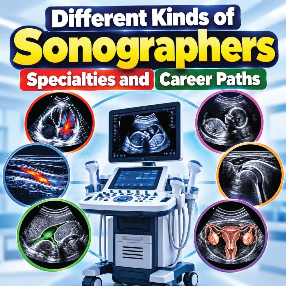
Ultrasound is used in many areas of modern healthcare, and the professionals who perform these examinations can specialize in very different parts of the body. From examining the heart and blood vessels to imaging the abdomen, breast tissue, reproductive system, or developing fetus, sonographers play an important role in producing diagnostic images for healthcare teams.
The term sonographer covers a wide range of specialties. An abdominal sonographer may focus on internal organs, while a cardiac sonographer performs echocardiograms to evaluate the heart. Vascular sonographers study blood flow, and obstetric and gynecologic sonographers work with pregnancy and reproductive health imaging. Other specialists focus on breast, pediatric, or musculoskeletal ultrasound.
Understanding the different kinds of sonographers is useful whether you are considering a career in diagnostic medical sonography or simply want to understand how ultrasound professionals differ. Each specialty involves its own anatomy, examination techniques, patient populations, and areas of clinical knowledge.
In this guide, we’ll explore the major sonography specialties, what each type of sonographer does, the skills and education involved, and how these career paths differ.
What Are the Different Kinds of Sonographers?
Sonography is a broad healthcare profession rather than a single type of job. Sonographers use ultrasound equipment to create images of organs, tissues, blood vessels, and other structures inside the body. The images and related information are then used by physicians and other qualified healthcare professionals to evaluate patients and help diagnose or manage medical conditions.
Because different parts of the body require different imaging techniques, sonographers can develop expertise in specific clinical areas. Some focus on the abdomen, while others work with the heart, blood vessels, breasts, musculoskeletal system, or reproductive system.
Common kinds of sonographers include:
- Abdominal sonographers
- Breast sonographers
- Cardiac sonographers or echocardiographers
- Vascular sonographers
- Obstetric and gynecologic sonographers
- Pediatric sonographers
- Musculoskeletal sonographers
- Fetal and pediatric cardiac sonographers
The exact scope of practice, credentials, and job titles can vary by employer, location, and professional certification. The Society of Diagnostic Medical Sonography describes sonography as a multispecialty profession that includes abdominal, breast, cardiac, musculoskeletal, obstetric/gynecologic, pediatric, venous, and vascular areas.
Abdominal Sonographers
Abdominal sonographers specialize in ultrasound examinations of the abdomen and nearby organs. Their work can involve imaging structures such as the liver, gallbladder, kidneys, pancreas, and spleen.
During an examination, the sonographer positions the patient, selects the appropriate transducer, adjusts imaging settings, and captures the views required for the examination.
What Does an Abdominal Sonographer Do?
An abdominal sonographer may perform examinations for patients being evaluated for symptoms or conditions involving abdominal organs.
Typical responsibilities include:
- Preparing the patient for the examination
- Explaining the procedure and answering questions
- Positioning the patient appropriately
- Obtaining diagnostic ultrasound images
- Checking images for quality and adequate coverage
- Recording measurements and relevant observations
- Communicating examination information to the interpreting physician
- Assisting with ultrasound-guided procedures when within the individual’s training and scope
Abdominal sonography requires careful attention to anatomy because structures can appear differently depending on the patient’s position, breathing, body composition, and other factors.
Breast Sonographers
Breast sonographers specialize in imaging breast tissue. Breast ultrasound can be used to further evaluate findings identified through physical examination or other breast imaging.
According to the U.S. Bureau of Labor Statistics, breast sonographers may help evaluate cysts and tumors and can assist with procedures involving ultrasound guidance.
What Is Breast Sonography Like?
Breast ultrasound is different from simply performing a general scan. The sonographer must understand breast anatomy and recognize the imaging characteristics that need careful documentation.
The examination may involve:
- Positioning the patient correctly
- Applying ultrasound gel
- Systematically scanning the breast tissue
- Imaging areas of concern
- Taking precise measurements
- Documenting relevant findings
- Providing images for physician interpretation
Breast sonographers also need strong communication skills because patients may feel anxious when an examination is being performed to investigate a breast abnormality.
Cardiac Sonographers and Echocardiographers
Cardiac sonographers, commonly called echocardiographers, specialize in ultrasound imaging of the heart. They examine structures such as the heart chambers, valves, and related vessels.
An echocardiogram can provide information about cardiac anatomy and function. Depending on the examination, cardiac sonographers may perform studies while a patient is resting or following physical activity.
What Does a Cardiac Sonographer Do?
Cardiac sonography requires a detailed understanding of cardiovascular anatomy and physiology.
A cardiac sonographer may:
- Perform echocardiographic examinations
- Obtain images of cardiac chambers and valves
- Capture measurements required for the examination
- Evaluate image quality
- Work with physicians and cardiology teams
- Perform specialized cardiac studies within their training
- Assist with examinations of fetal or pediatric hearts when appropriately trained
Cardiac sonography can therefore involve different patient groups, including adults, children, and unborn babies.
Vascular Sonographers
Vascular sonographers focus on blood vessels and blood flow. Their examinations can help physicians evaluate conditions involving arteries and veins.
Unlike a basic anatomical ultrasound, vascular studies often combine imaging with information about blood movement. Doppler ultrasound is particularly important in this area because it can provide information about the direction and characteristics of blood flow.
What Does a Vascular Sonographer Examine?
Depending on the clinical setting and scope of practice, vascular sonographers may examine areas such as:
- Carotid arteries
- Peripheral arteries
- Peripheral veins
- Abdominal vessels
- Blood flow in the extremities
Vascular sonographers may help identify findings associated with problems such as blocked arteries or blood clots.
Accuracy is particularly important because the images and measurements can contribute to decisions about further diagnostic testing or treatment.
Obstetric and Gynecologic Sonographers
Obstetric and gynecologic sonographers specialize in ultrasound examinations involving pregnancy and the female reproductive system.
Obstetric sonography can be used throughout pregnancy to evaluate fetal development and anatomy, while gynecologic examinations can assess reproductive organs and related structures.
What Does an OB/GYN Sonographer Do?
Depending on the examination, an OB/GYN sonographer may perform imaging involving:
- The uterus
- Ovaries
- Endometrium
- Developing fetus
- Placenta
- Amniotic environment
- Pelvic structures
Pregnancy examinations require both technical skill and sensitive patient communication. Patients may have questions or concerns throughout the examination, so the sonographer needs to explain the process appropriately while staying within the professional scope of the role.
Pediatric Sonographers
Pediatric sonographers specialize in imaging infants, children, and, in some settings, newborn patients. Their examinations may involve conditions associated with premature birth, congenital abnormalities, or other pediatric medical concerns.
Why Is Pediatric Sonography Different?
Children are not simply smaller versions of adult patients. Their anatomy, communication needs, movement, and ability to cooperate with an examination can differ considerably.
Pediatric sonographers may need to:
- Adapt communication to the child’s age
- Help children remain comfortable
- Work with parents or caregivers
- Use appropriate examination techniques
- Obtain diagnostic images while minimizing unnecessary delays
- Work closely with pediatric physicians and other healthcare professionals
Patience and communication are especially valuable in this specialty.
Also Read:
Musculoskeletal Sonographers
Musculoskeletal sonographers use ultrasound to examine muscles, tendons, ligaments, joints, and related soft tissues.
This specialty can be useful for evaluating certain sports injuries, tendon problems, joint abnormalities, and other conditions involving superficial musculoskeletal structures.
What Can Musculoskeletal Sonographers Image?
Depending on the clinical environment, examinations can involve:
- Tendons
- Muscles
- Ligaments
- Joints
- Soft-tissue structures
The U.S. Bureau of Labor Statistics notes that musculoskeletal sonographers may also assist with ultrasound-guided injections or surgical procedures when applicable.
Musculoskeletal imaging can require the sonographer to understand anatomy in motion. Some examinations may involve dynamic scanning, where a structure is observed while the patient moves in a controlled manner.
Fetal and Pediatric Cardiac Sonographers
Cardiac sonography itself contains additional areas of specialization. Some professionals focus on adult echocardiography, while others develop expertise in fetal or pediatric cardiac imaging.
Fetal echocardiography focuses on the developing baby’s heart. Pediatric echocardiography focuses on infants and children.
These areas require specialized knowledge because cardiac anatomy and physiology change significantly throughout development.
Professional credentialing organizations recognize several cardiac and related specialties. ARDMS credentials and specialty areas include adult echocardiography, fetal echocardiography, pediatric echocardiography, and pediatric sonography, among others.
Also Read:
Sonography Specialties at a Glance
The following table provides a simple overview of the major types of sonographers.
| Sonography Specialty | Primary Area Examined | Common Focus |
|---|---|---|
| Abdominal | Abdominal organs | Liver, kidneys, gallbladder, pancreas, spleen |
| Breast | Breast tissue | Cysts, masses, tissue abnormalities |
| Cardiac | Heart | Chambers, valves, cardiac function |
| Vascular | Blood vessels | Arteries, veins, blood flow |
| OB/GYN | Reproductive system and pregnancy | Pelvis, pregnancy, fetal development |
| Pediatric | Infants and children | Pediatric organs and conditions |
| Musculoskeletal | Muscles and joints | Tendons, ligaments, muscles, joints |
| Fetal cardiac | Fetal heart | Developing cardiac anatomy |
| Pediatric cardiac | Children’s hearts | Pediatric cardiac anatomy and function |
The boundaries between specialties can vary between employers and professional organizations, and some sonographers develop competencies in more than one area.
How Sonographers Choose a Specialty
Choosing a sonography specialty is often influenced by education, clinical experience, employer needs, professional interests, and certification pathways.
A student may first develop general sonography knowledge and then gain clinical experience in a particular area. Some educational programs provide focused instruction in specialties such as abdominal or breast sonography as part of the curriculum.
Factors That Can Influence the Choice
Someone considering a sonography career may think about:
- Which anatomy they enjoy studying
- Whether they prefer adult or pediatric patients
- Interest in pregnancy and women’s health
- Interest in cardiovascular medicine
- Interest in vascular anatomy and blood flow
- Interest in sports injuries and musculoskeletal anatomy
- Desired clinical environment
- Available training programs
- Certification requirements
- Local employer opportunities
There is no single path that fits every future sonographer. Specialty opportunities depend heavily on the healthcare system, employer, and region.
Also Read:
What Skills Do Different Sonographers Need?
Although specialties differ, many core skills are shared across the profession.
Diagnostic medical sonographers need to operate sophisticated ultrasound equipment while simultaneously observing the resulting images. They also need to position patients correctly and obtain the views required for clinical interpretation.
Technical and Clinical Skills
Important skills include:
- Knowledge of anatomy
- Understanding of ultrasound physics
- Image optimization
- Hand-eye coordination
- Accurate measurement
- Patient positioning
- Equipment operation
- Attention to detail
- Documentation
- Communication with healthcare professionals
The physical side of the job also matters. Sonographers may stand for long periods and may need to assist with positioning or moving patients who require help.
Education and Certification for Sonographers
In the United States, diagnostic medical sonographers typically enter the profession with at least an associate degree or a postsecondary certificate, although bachelor’s degree programs are also available. Sonography education commonly includes anatomy, medical terminology, applied sciences, specialized ultrasound instruction, and clinical experience.
Certification and licensing requirements depend on the location and employer. Some states have licensing requirements, while employers may require or prefer professional certification.
Professional credentials can also be specialty-specific. For example, ARDMS recognizes credentials and specialty areas covering diagnostic medical sonography, cardiac sonography, vascular technology, and musculoskeletal sonography.
Students should therefore check the requirements applicable to the state, country, school, and employers they are considering.
Where Do Different Sonographers Work?
Sonographers work in several healthcare environments. Hospitals represent a major employment setting, but physician offices, diagnostic laboratories, outpatient centers, and other healthcare facilities also employ sonography professionals.
| Work Setting | Examples of Sonography Work |
|---|---|
| Hospitals | General, emergency, inpatient, and specialty imaging |
| Physician offices | Outpatient examinations |
| Diagnostic laboratories | Scheduled imaging studies |
| Outpatient centers | Specialized diagnostic examinations |
| Specialty healthcare facilities | Focused clinical imaging |
The daily experience can differ considerably between settings. A hospital-based sonographer may encounter a wider variety of patients and urgent examinations, while an outpatient facility may have a more predictable schedule.
Are All Sonographers Called Ultrasound Technicians?
The terms “sonographer” and “ultrasound technician” are often used interchangeably in everyday conversation, but professional titles can be more specific.
A diagnostic medical sonographer is a healthcare professional trained to perform ultrasound examinations and acquire diagnostic images. Within that broader profession, someone may identify as an abdominal sonographer, cardiac sonographer, vascular technologist, breast sonographer, or another specialty professional.
The distinction matters because specialization reflects differences in the examinations performed, anatomy studied, and training or credentials held.
What Is the Career Outlook for Sonographers?
The U.S. Bureau of Labor Statistics reports that diagnostic medical sonographers had about 92,200 jobs in 2025 and projects employment to reach approximately 105,100 by 2035. The occupation is projected to grow 14% from 2025 to 2035, with about 6,000 openings per year on average.
The same BLS data reports a median annual wage of $96,590 for diagnostic medical sonographers in May 2025. Pay varies by industry, location, experience, specialty, and employer, so national figures should not be treated as an individual salary guarantee.
The expansion of ultrasound applications across medical specialties is one factor identified by BLS in its employment outlook.
Which Type of Sonographer Is Right for You?
Different kinds of sonographers work with different anatomy, patient populations, and clinical questions. Someone interested in cardiovascular anatomy may explore cardiac sonography, while a person interested in pregnancy may prefer obstetric imaging. Another student may enjoy musculoskeletal anatomy, pediatric care, vascular studies, or abdominal imaging.
A useful way to compare specialties is to consider four questions:
- What part of the body interests you most?
- Which patient population do you prefer?
- What type of clinical environment suits you?
- Which education and certification pathways are available to you?
Clinical exposure is particularly valuable because the day-to-day reality of a specialty can be different from what a classroom description suggests.
Final Thoughts on the Different Kinds of Sonographers
There are many different kinds of sonographers, and each specialty uses ultrasound technology for a particular clinical purpose. Abdominal, breast, cardiac, vascular, OB/GYN, pediatric, and musculoskeletal sonographers all share the fundamentals of ultrasound imaging while developing additional knowledge for their chosen area.
For someone considering sonography as a career, understanding these specialties can make education and career planning easier. The best starting point is to learn about the examinations, patient populations, training requirements, and certification expectations associated with each specialty.
Sonography continues to be a diverse field because ultrasound can be applied across many areas of medicine. That variety allows professionals to build careers around different interests while contributing important diagnostic information to the healthcare team.

Zak is a dedicated medical and career writer specializing in sonography, healthcare education, and professional development. Through SonographerSalary.com, he shares in-depth insights on sonographer salaries, education pathways, and career tips to help readers build successful futures in medical imaging. His content combines accuracy with practical, easy-to-understand guidance, empowering students and professionals to make confident, informed career decisions.