
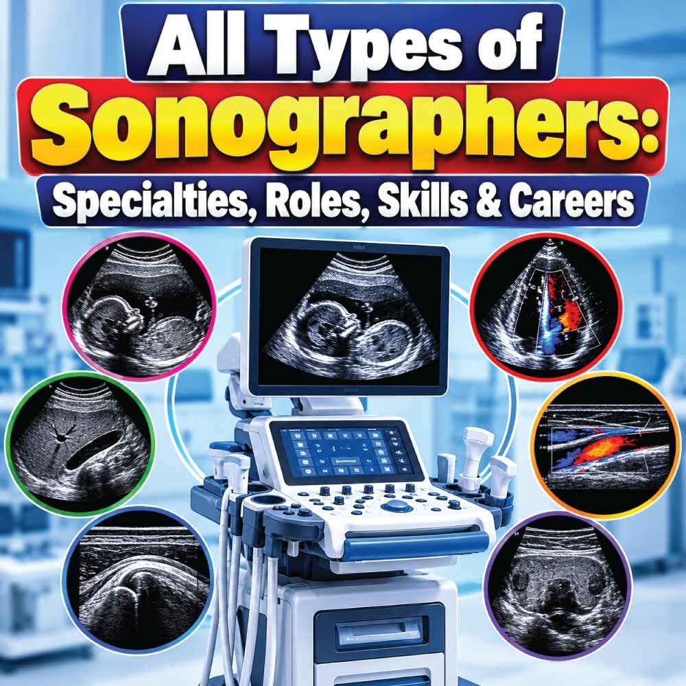
Sonography is a diverse healthcare profession with many different specialties, each focused on a particular part of the body, type of examination, or patient group. While all sonographers use ultrasound technology to create diagnostic images, their daily responsibilities can vary considerably depending on their area of specialization.
There are abdominal sonographers who examine internal organs, vascular sonographers who assess blood vessels and blood flow, cardiac sonographers who perform echocardiograms, and OB/GYN sonographers who specialize in pregnancy and reproductive imaging. Other professionals focus on breast, pediatric, musculoskeletal, or fetal cardiac ultrasound.
Understanding the types of sonographers can be helpful if you are considering a career in diagnostic medical sonography or simply want to know how ultrasound professionals differ. Each specialty requires a combination of technical knowledge, anatomy skills, patient care, and hands-on scanning experience.
In this guide, we will explore the major sonography specialties, what each type of sonographer does, the anatomy they examine, common credentials, required skills, and how to choose a specialty that matches your interests and career goals.
What Are the Different Types of Sonographers?
Sonography is much broader than the general idea of performing an ultrasound scan. Sonographers can specialize in different areas of the body, patient populations, and types of examinations. Their work may involve abdominal organs, blood vessels, the heart, pregnancy, breast tissue, muscles and tendons, or pediatric patients.
The Society of Diagnostic Medical Sonography (SDMS) describes sonography as a multispecialty profession that includes abdominal, breast, cardiac, musculoskeletal, obstetric and gynecologic, pediatric, venous, and vascular practice areas.
A sonographer uses an ultrasound transducer to send high-frequency sound waves into the body and produce diagnostic images or other information. Those images are then provided to the interpreting physician or other authorized healthcare professional.
The major types of sonographers include:
- Abdominal sonographers
- Obstetric and gynecologic sonographers
- Cardiac or echocardiography sonographers
- Vascular sonographers
- Breast sonographers
- Pediatric sonographers
- Musculoskeletal sonographers
- Fetal echocardiography specialists
- Other specialized and emerging ultrasound professionals
The exact title, scope of practice, credential, and licensing requirements can vary by location and employer.
Abdominal Sonographers
Abdominal sonographers specialize in imaging structures within the abdomen and related areas. Their examinations can involve organs such as the liver, gallbladder, pancreas, spleen, kidneys, abdominal aorta, and other structures.
Abdominal ultrasound is commonly used when physicians need information about abdominal pain, swelling, suspected disease, masses, fluid, or abnormalities involving abdominal organs.
What Does an Abdominal Sonographer Do?
An abdominal sonographer prepares the patient, selects appropriate equipment and transducers, obtains diagnostic images, and adjusts scanning techniques when necessary.
Depending on the examination, the sonographer may evaluate:
- Liver
- Gallbladder and bile ducts
- Pancreas
- Kidneys
- Spleen
- Abdominal aorta
- Retroperitoneal structures
- Superficial abdominal structures
The ARDMS abdominal examination content includes evaluation of multiple abdominal and related structures, demonstrating how broad this specialty can be.
Abdominal sonography is therefore not simply one standard scan. The examination is adapted to the patient’s symptoms, medical history, physician’s order, and the structures being investigated.
Obstetric and Gynecologic Sonographers
Obstetric and gynecologic, often called OB/GYN, sonography focuses on the female reproductive system and pregnancy-related imaging.
Obstetric examinations can be performed at different stages of pregnancy and may provide information about fetal development, anatomy, position, and other clinical findings.
Gynecologic examinations can involve structures such as the uterus, ovaries, cervix, and surrounding pelvic anatomy.
What Makes OB/GYN Sonography Different?
This specialty requires both technical scanning skills and the ability to communicate respectfully with patients during examinations that can be personal or emotionally significant.
The sonographer may need to:
- Explain the examination process
- Position the patient appropriately
- Obtain required images and measurements
- Evaluate fetal or pelvic anatomy according to the examination protocol
- Document relevant images and information
- Maintain privacy and professional communication
OB/GYN is one of the recognized specialty areas within diagnostic medical sonography and is also an established ARDMS specialty.
Cardiac Sonographers and Echocardiographers
Cardiac sonographers specialize in imaging the heart. They are commonly known as echocardiographers because the examinations they perform are called echocardiograms or echocardiography studies.
Instead of concentrating primarily on abdominal organs or pregnancy, cardiac sonographers examine cardiac anatomy and function.
What Does a Cardiac Sonographer Examine?
A cardiac examination can provide information about:
- Heart chambers
- Heart valves
- Blood flow
- Cardiac motion
- Heart muscle function
- Certain structural abnormalities
- Prosthetic valves and cardiac devices
Doppler ultrasound is particularly important in cardiac imaging because it can help evaluate blood flow and hemodynamic information.
Cardiac sonography includes several areas, including adult echocardiography, pediatric echocardiography, and fetal echocardiography. ARDMS recognizes cardiac credentials and specialty examinations within its credentialing system.
Adult Echocardiography
Adult echocardiographers primarily examine the hearts of adult patients. Their work may take place in hospitals, outpatient cardiac facilities, cardiology practices, and other healthcare environments.
Pediatric Echocardiography
Pediatric cardiac sonographers work with infants, children, and adolescents. The examinations can require specialized technical knowledge as well as communication techniques suited to younger patients and their families.
Vascular Sonographers
Vascular sonographers specialize in examining blood vessels and blood flow. Their work frequently combines conventional ultrasound imaging with Doppler techniques.
They may evaluate arteries and veins in different areas of the body, depending on the examination requested.
What Does a Vascular Sonographer Do?
Vascular examinations may assess areas such as:
- Carotid arteries
- Peripheral arteries
- Peripheral veins
- Abdominal or visceral vessels
- Other vascular structures
Doppler technology allows the sonographer to evaluate blood movement and related characteristics rather than simply producing a static anatomical image.
Vascular technology is recognized as a distinct area of sonography, and vascular sonography is included among the specialty areas represented by professional organizations and credentialing pathways.
Because vascular examinations can require detailed measurements and careful technique, vascular sonographers need a strong understanding of ultrasound physics, anatomy, Doppler principles, and examination protocols.
Breast Sonographers
Breast sonographers perform ultrasound examinations of breast tissue. Breast ultrasound may be used alongside other forms of breast imaging or as part of a diagnostic evaluation.
The examination can focus on a particular area of concern or involve a broader assessment depending on the clinical situation.
What Can Breast Ultrasound Evaluate?
Breast sonography can help characterize findings such as:
- Cysts
- Solid masses
- Changes in breast tissue
- Areas identified during another examination
- Superficial structures in the breast
The sonographer’s job is to obtain technically appropriate images and document the findings required by the examination protocol. Interpretation of diagnostic findings is performed by the appropriate physician or qualified healthcare professional.
Breast sonography is recognized as a specialty within professional sonography education and credentialing resources.
Pediatric Sonographers
Pediatric sonographers specialize in ultrasound examinations for children. Pediatric imaging can involve many of the same anatomical regions examined in adults, but the approach may be quite different.
Children can have different anatomical characteristics, developmental stages, communication needs, and responses to medical environments.
What Skills Are Important in Pediatric Sonography?
A pediatric sonographer needs strong technical skills, but patient interaction is equally important.
Useful abilities include:
- Explaining procedures in age-appropriate language
- Keeping children comfortable and cooperative
- Working with parents or guardians
- Adjusting scanning techniques for smaller anatomy
- Maintaining a calm clinical environment
- Obtaining diagnostic-quality images efficiently
Pediatric sonography is identified as a distinct specialty in professional education resources, while pediatric cardiac sonography is treated as a separate cardiac specialty.
Also Read:
Fetal Echocardiography Sonographers
Fetal echocardiography is a highly specialized area involving ultrasound evaluation of the developing fetal heart.
While general obstetric sonography can include assessment of fetal anatomy, fetal echocardiography focuses specifically on cardiac structures and function.
How Is Fetal Echocardiography Different From Routine Obstetric Ultrasound?
A routine obstetric examination may evaluate many aspects of pregnancy and fetal development. A fetal echocardiogram is more specifically directed toward the fetal cardiovascular system.
The examination can require detailed knowledge of:
- Fetal cardiac anatomy
- Cardiac chambers
- Valves
- Blood flow
- Cardiac motion
- Congenital cardiac abnormalities
Fetal echocardiography is recognized as a specialized cardiac sonography area by professional organizations and credentialing resources.
Musculoskeletal Sonographers
Musculoskeletal, or MSK, sonographers focus on muscles, tendons, ligaments, joints, nerves, and other superficial soft tissues.
This specialty can be particularly useful in sports medicine, orthopedics, rehabilitation, and other clinical settings where dynamic assessment of soft tissue is important.
Ultrasound can be used to evaluate structures such as tendons and muscles while they move, providing information that may complement other imaging methods.
What Can MSK Sonography Examine?
Depending on the clinical indication, examinations can involve:
- Rotator cuff tendons
- Muscles
- Ligaments
- Peripheral nerves
- Tendons of the hand and wrist
- Soft-tissue abnormalities
- Joint-related structures
Musculoskeletal sonography requires detailed anatomical knowledge and excellent hand-eye coordination because the sonographer may need to manipulate the transducer while observing structures dynamically.
Also Read:
General Sonographers vs Specialized Sonographers
Not every sonographer works exclusively within one narrow specialty. Some professionals develop broader skills and perform examinations across multiple areas.
The distinction between a general sonographer and a specialist often depends on education, credentials, employer requirements, clinical setting, and local regulations.
| Sonographer Type | Primary Focus | Common Examination Areas |
|---|---|---|
| General/Abdominal | Internal organs | Liver, kidneys, gallbladder, pancreas |
| OB/GYN | Pregnancy and reproductive anatomy | Fetus, uterus, ovaries |
| Cardiac | Heart | Chambers, valves, cardiac function |
| Vascular | Blood vessels | Arteries, veins, blood flow |
| Breast | Breast tissue | Masses, cysts, tissue abnormalities |
| Pediatric | Children | Age-specific ultrasound examinations |
| MSK | Muscles and soft tissues | Tendons, muscles, nerves |
| Fetal Cardiac | Fetal heart | Fetal cardiac anatomy and function |
The categories can overlap. For example, a professional may have expertise in both abdominal and breast imaging, or combine obstetric and gynecologic examinations in an OB/GYN practice.
Also Read:
How Sonography Specialties Differ
Although every sonographer uses ultrasound technology, the day-to-day work can be very different from one specialty to another.
| Specialty | Main Anatomy | Important Technical Skills | Typical Patient Group |
|---|---|---|---|
| Abdominal | Abdominal organs | Gray-scale imaging, anatomy | Mostly adults |
| OB/GYN | Reproductive system and fetus | Obstetric protocols, measurements | Pregnant patients and adults |
| Cardiac | Heart | Doppler, cardiac imaging | Adults or children |
| Vascular | Arteries and veins | Spectral/color Doppler | Adults and other patients |
| Breast | Breast tissue | Tissue characterization | Mostly adults |
| Pediatric | Various anatomy | Pediatric scanning techniques | Children |
| MSK | Muscles, tendons, nerves | Dynamic scanning | Adults and athletes |
| Fetal Cardiac | Fetal heart | Detailed cardiac imaging | Pregnant patients |
These differences are one reason sonography is considered a multispecialty profession rather than a single uniform job.
What All Types of Sonographers Have in Common
Despite their different specialties, sonographers share many fundamental responsibilities.
They must understand ultrasound physics, anatomy, patient care, equipment operation, image optimization, documentation, and professional communication.
The ARDMS Sonography Principles and Instrumentation examination includes areas such as performing ultrasound examinations, managing transducers, optimizing images, applying Doppler concepts, and clinical safety and quality assurance.
Core Skills Used Across Sonography
A sonographer commonly needs to be able to:
- Select an appropriate transducer
- Position patients safely and comfortably
- Adjust imaging settings
- Optimize diagnostic images
- Apply Doppler techniques when required
- Recognize technical limitations and artifacts
- Follow examination protocols
- Communicate professionally with patients
- Document required images and information
- Maintain appropriate infection-control and safety practices
Ergonomics is also important because sonographers can perform repetitive scanning movements for long periods. SDMS includes ergonomically appropriate scanning techniques within the professional scope of practice.
What Credentials Do Sonographers Use?
Credentials depend on the specialty and certifying organization. In the United States, ARDMS offers several major sonography credentials, including RDMS, RDCS, and RVT, with specialty examinations associated with particular areas of practice.
Some commonly encountered credentials include:
| Credential | Meaning | Broad Area |
|---|---|---|
| RDMS | Registered Diagnostic Medical Sonographer | Diagnostic sonography |
| RDCS | Registered Diagnostic Cardiac Sonographer | Cardiac sonography |
| RVT | Registered Vascular Technologist | Vascular sonography |
| RMSKS | Registered Musculoskeletal Sonographer | Musculoskeletal sonography |
A credential should not be confused with a job title. Employers and jurisdictions can have their own education, certification, registration, and licensing requirements.
Anyone considering this career should check the requirements that apply where they intend to work.
How to Choose a Sonography Specialty
Choosing a specialty is partly a matter of personal interest and partly a practical career decision. Students should consider what type of anatomy and patient care they enjoy before committing to a particular pathway.
Consider the Type of Patients You Want to Work With
If you enjoy working with expectant parents and pregnancy-related imaging, OB/GYN may be interesting. If you prefer cardiovascular anatomy, echocardiography may be a better area to investigate.
People who enjoy working with children may prefer pediatric sonography, while those interested in sports injuries and anatomy in motion may find MSK imaging appealing.
Consider the Type of Imaging You Enjoy
Some specialties rely heavily on Doppler, while others focus more on detailed anatomical imaging.
For example:
- Vascular sonography uses Doppler extensively.
- Cardiac sonography combines anatomy, motion, and blood-flow assessment.
- MSK sonography often involves dynamic evaluation.
- Abdominal sonography requires broad knowledge of internal anatomy.
- OB/GYN requires specialized pregnancy and pelvic imaging knowledge.
Researching clinical rotations and observing experienced sonographers can help students understand what each specialty is actually like in practice.
Can a Sonographer Have More Than One Specialty?
Yes. Sonographers can develop competencies in multiple specialty areas, although the exact opportunities depend on their education, clinical experience, credentials, employer, and local regulations.
Professional education resources list multiple specialty pathways, including abdomen, adult cardiac, breast, fetal cardiac, OB/GYN, pediatric, pediatric cardiac, and vascular sonography.
Having experience across specialties can allow a professional to work in different clinical environments. However, gaining an additional specialty generally requires appropriate education, supervised clinical experience, competency, and any applicable credentialing.
Where Do Different Types of Sonographers Work?
Sonographers can work in a variety of healthcare environments rather than only traditional hospitals.
Possible workplaces include:
- Hospitals
- Outpatient imaging centers
- Physician offices
- Cardiology practices
- Vascular laboratories
- Women’s health clinics
- Pediatric hospitals
- Orthopedic and sports medicine facilities
- Specialty diagnostic centers
The work environment often depends on the sonographer’s specialty. For example, cardiac sonographers are commonly associated with cardiac departments and cardiology practices, while vascular sonographers may work in dedicated vascular laboratories or hospitals.
What Is the Future of Sonography Specialization?
Ultrasound continues to expand into different clinical applications. Professional standards already identify established specialties as well as emerging clinical areas.
Advances in equipment, Doppler technology, three-dimensional imaging, portable ultrasound, and ultrasound-guided procedures are broadening the ways ultrasound can be used.
However, technology does not eliminate the need for skilled sonographers. Producing useful diagnostic images requires more than operating a machine. The professional must understand anatomy, recognize limitations, select appropriate techniques, communicate with patients, and obtain the information needed for clinical interpretation.
Final Thoughts on the Types of Sonographers
There is no single type of sonographer. The profession includes specialists who focus on abdominal imaging, pregnancy, women’s health, cardiac imaging, vascular studies, breast ultrasound, pediatrics, musculoskeletal imaging, and fetal cardiac examinations.
Each specialty uses the same fundamental ultrasound technology but applies it to different anatomy, patients, and clinical questions.
For students considering sonography as a career, understanding these specialties is an important first step. The right pathway depends on interests, strengths, education, clinical opportunities, credentialing requirements, and the type of patients and examinations a person wants to work with.
Most importantly, specialization does not change the central responsibility of the profession: obtaining high-quality diagnostic information while providing safe, respectful, and patient-centered care.

Zak is a dedicated medical and career writer specializing in sonography, healthcare education, and professional development. Through SonographerSalary.com, he shares in-depth insights on sonographer salaries, education pathways, and career tips to help readers build successful futures in medical imaging. His content combines accuracy with practical, easy-to-understand guidance, empowering students and professionals to make confident, informed career decisions.