
Ultrasound is one of the most widely used imaging techniques in modern medicine, known for being safe, painless, and highly effective in viewing internal structures of the human body. Unlike X-rays or CT scans, it does not use radiation, which makes it especially valuable for pregnant women, children, and patients who require repeated imaging.
Ultrasound works by using high-frequency sound waves that bounce off tissues and organs inside the body. These reflected sound waves are then converted into real-time images on a screen. Because of its versatility and safety, ultrasound has become a core diagnostic tool in hospitals, clinics, and emergency care units worldwide.
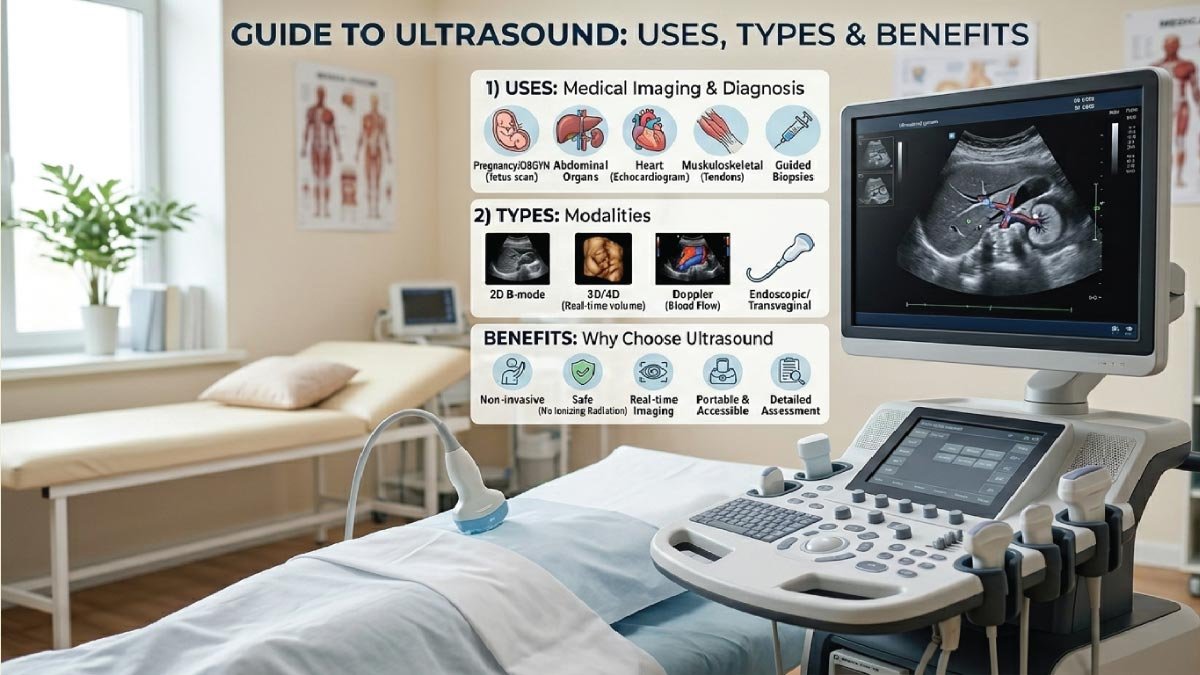
In this guide, we will explore what ultrasound is, how it works, its major types, common uses, and the key benefits that make it such an important medical technology.
What Is Ultrasound?
Ultrasound is a diagnostic imaging method that uses sound waves above the range of human hearing (typically above 20,000 Hz). In medical applications, these sound waves are much higher—usually between 2 to 18 megahertz.
A device called a transducer sends sound waves into the body. When these waves hit organs, fluids, or tissues, they bounce back at different speeds and intensities. The machine then processes these returning signals to create a live image.
This allows doctors to see movement inside the body, such as:
- A beating heart
- Blood flowing through vessels
- A developing fetus
- Organ structure and abnormalities
Because the images are real-time, ultrasound is also used for guiding medical procedures like needle biopsies or injections.
How Does Ultrasound Work?
Ultrasound works by sending high-frequency sound waves into the body and capturing their echoes to create real-time images. These waves interact with different tissues, producing signals that are converted into detailed visuals for medical diagnosis. The process is fast, safe, and completely non-invasive.
The working process of ultrasound can be broken down into simple steps:
1. Sound Wave Generation
The transducer emits high-frequency sound waves into the body using a special gel that improves contact with the skin. This gel removes air gaps and ensures smooth transmission of sound waves, allowing accurate penetration into internal tissues for clear imaging results.
2. Wave Reflection
When sound waves travel through different tissues, they bounce back differently depending on density. For example:
- Fluid-filled areas appear dark
- Dense tissues appear lighter
These variations help doctors distinguish between normal and abnormal structures inside the body, making diagnosis more precise and visually understandable.
3. Signal Conversion
The returning echoes are collected by the transducer and sent to a computer system. These echoes carry detailed information about tissue shape, density, and structure, which the system processes to form meaningful data for creating diagnostic images.
4. Image Formation
The system processes these echoes into visual images displayed on a monitor in real time. This allows doctors to observe organs, blood flow, and movements instantly, helping in quick diagnosis and immediate medical decision-making during examinations or procedures.
This process happens extremely fast, allowing doctors to see internal body functions as they occur.
Types of Ultrasound
Ultrasound technology has developed into several specialized types, each designed for specific medical applications. These different forms help doctors examine organs, monitor pregnancies, study blood flow, and evaluate internal conditions with greater accuracy and detail.
1. 2D Ultrasound
This is the most common type and produces flat, two-dimensional images. It provides basic but essential visual information about internal organs and fetal development, making it widely used in routine medical checkups and pregnancy monitoring.
Common uses:
- Monitoring fetal development
- Examining abdominal organs
- Checking kidney or liver conditions
2. 3D Ultrasound
3D ultrasound captures multiple images from different angles and combines them into a three-dimensional view. It provides a more realistic and detailed representation of body structures, improving diagnostic accuracy and helping parents see clearer images during pregnancy scans.
Benefits include:
- Clearer image of fetal face and body
- Better detection of physical abnormalities
- Improved surgical planning
3. 4D Ultrasound
4D ultrasound adds the element of time to 3D images, showing movement in real time. It allows doctors to observe dynamic activities inside the body, such as fetal movement or heart activity, with enhanced clarity and detail.
Common uses:
- Watching fetal movements
- Assessing heart function
- Studying organ motion
4. Doppler Ultrasound
Doppler ultrasound measures blood flow in veins and arteries by analyzing sound wave changes caused by moving blood cells. It is essential for evaluating circulation problems and detecting cardiovascular issues early.
It helps detect:
- Blood clots
- Blocked arteries
- Poor circulation
- Heart valve problems
This type is especially important in cardiovascular diagnosis.
5. Echocardiogram
An echocardiogram is a specialized ultrasound focused on the heart, providing detailed images of its structure and function. It helps doctors understand how effectively the heart is pumping and whether any abnormalities are present.
It helps evaluate:
- Heart structure
- Pumping efficiency
- Valve function
Doctors often use it to diagnose heart disease or monitor ongoing conditions.
6. Transvaginal Ultrasound
This type uses a specially designed probe inserted into the vaginal canal for clearer pelvic images. It provides close-range imaging, offering better detail of reproductive organs compared to traditional abdominal scans.
It is commonly used for:
- Early pregnancy scanning
- Fertility evaluation
- Detecting uterine or ovarian conditions
7. Abdominal Ultrasound
This scan focuses on abdominal organs like the liver, gallbladder, pancreas, and kidneys. It helps doctors identify abnormalities in internal organs and is commonly used for diagnosing digestive and urinary system issues.
It helps detect:
- Gallstones
- Liver disease
- Kidney stones
- Abnormal growths
Also Read:
Uses of Ultrasound in Medicine
Ultrasound is used across nearly all medical fields due to its flexibility and safety.
1. Pregnancy and Obstetrics
One of the most well-known uses of ultrasound is monitoring pregnancy. It helps track fetal growth, detect abnormalities, and confirm gestational age.
Doctors use it to:
- Confirm pregnancy
- Monitor fetal heartbeat
- Check baby position
- Estimate due date
2. Cardiology
In heart care, ultrasound helps doctors evaluate heart health and detect cardiovascular diseases.
It is used for:
- Checking heart valves
- Measuring blood flow
- Identifying heart enlargement
- Detecting fluid around the heart
3. Abdominal Diagnosis
Ultrasound is widely used to examine internal organs for disease or damage.
It can detect:
- Tumors
- Infections
- Organ enlargement
- Stones in kidneys or gallbladder
4. Musculoskeletal Imaging
Doctors use ultrasound to study muscles, tendons, and joints.
It helps diagnose:
- Sports injuries
- Muscle tears
- Joint inflammation
- Ligament damage
5. Emergency Medicine
In emergency rooms, ultrasound provides quick diagnostic information.
It is used to:
- Detect internal bleeding
- Check organ damage after trauma
- Guide emergency procedures
6. Guided Medical Procedures
Ultrasound helps doctors perform procedures with precision.
Examples include:
- Needle biopsies
- Fluid drainage
- Injections
- Catheter placement
Also Read:
Limitations of Ultrasound
Although highly useful, ultrasound does have some limitations.
- It cannot pass through bone or air-filled organs like lungs
- Image quality depends on the operator’s skill
- Deep tissues may not be clearly visible
- It may not detect very small or early-stage abnormalities
Despite these limitations, it remains one of the most valuable diagnostic tools in medicine.
Future of Ultrasound Technology
Ultrasound technology continues to improve rapidly. Modern advancements include:
- AI-powered image analysis
- 3D and 4D high-definition imaging
- Wireless handheld ultrasound devices
- Improved Doppler accuracy
- Faster and more detailed scanning systems
These innovations are making ultrasound even more powerful, accessible, and accurate for doctors worldwide.
Conclusion
Ultrasound is a safe, effective, and versatile medical imaging technique that plays a vital role in modern healthcare. From pregnancy monitoring to heart disease detection, its applications are vast and essential.
With different types such as 2D, 3D, Doppler, and echocardiography, ultrasound provides doctors with detailed insights into the human body without using harmful radiation. Its benefits—such as safety, affordability, and real-time imaging—make it one of the most trusted diagnostic tools in the world.
As technology continues to advance, ultrasound will become even more precise and accessible, improving patient care and helping doctors diagnose conditions earlier and more accurately.
Also Read:

Zak is a dedicated medical and career writer specializing in sonography, healthcare education, and professional development. Through SonographerSalary.com, he shares in-depth insights on sonographer salaries, education pathways, and career tips to help readers build successful futures in medical imaging. His content combines accuracy with practical, easy-to-understand guidance, empowering students and professionals to make confident, informed career decisions.