
The curvilinear probe, also known as a convex probe, is one of the most widely used ultrasound transducers in diagnostic imaging. It is specifically designed to visualize deeper structures within the body and is commonly used for abdominal, obstetric, gynecological, and emergency ultrasound examinations.
Unlike the linear probe, which produces a rectangular image, the curvilinear probe creates a fan-shaped image that provides a wider field of view at greater depths. Its lower frequency allows ultrasound waves to penetrate deeper into the body, making it ideal for imaging organs and structures located far beneath the skin surface.
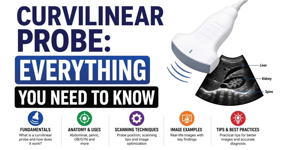
This guide explains everything you need to know about the curvilinear probe, including its design, frequency range, applications, advantages, limitations, and role in modern ultrasound imaging.
What Is a Curvilinear Probe?
A curvilinear probe is an ultrasound transducer that contains multiple piezoelectric crystal elements arranged along a curved surface.
These crystal elements emit and receive ultrasound waves to create diagnostic images of internal organs and tissues.
The curved footprint of the probe allows the ultrasound beam to spread outward as it travels deeper into the body, producing a broad field of view that is particularly useful for abdominal and pelvic examinations.
How Does a Curvilinear Probe Work?
The curvilinear probe works using the same ultrasound principles as other transducers.
The process includes:
- Electrical energy activates the crystal elements.
- The crystals produce ultrasound waves.
- Sound waves travel into the body.
- Internal structures reflect the waves.
- Echoes return to the probe.
- The ultrasound machine converts the echoes into images.
Because the crystal elements are arranged in a curved configuration, the resulting image expands outward, creating a sector or fan-shaped display.
Appearance of a Curvilinear Probe
The curvilinear probe has a distinctive curved footprint.
Characteristics include:
- Rounded scanning surface
- Convex-shaped face
- Wider field of view at depth
- Fan-shaped image display
- Larger footprint than phased array probes
Its design allows it to cover a large scanning area, making it ideal for evaluating abdominal organs and pregnancy-related structures.
Frequency Range of a Curvilinear Probe
Curvilinear probes typically operate at lower frequencies than linear probes.
Common frequency ranges include:
| Probe Type | Frequency Range |
|---|---|
| Standard Curvilinear Probe | 2–5 MHz |
| General Abdominal Probe | 3–6 MHz |
| Obstetric Probe | 2–7 MHz |
Lower frequencies provide:
- Greater penetration depth
- Better visualization of deep organs
- Wider scanning capability
However, image resolution is slightly lower compared to high-frequency probes.
Why Does Frequency Matter?
Ultrasound frequency determines image quality and penetration depth.
Low Frequency
Advantages:
- Deeper penetration
- Better visualization of abdominal organs
- Improved imaging in larger patients
Disadvantages:
- Reduced image detail
- Lower spatial resolution
High Frequency
Advantages:
- Sharper images
- Better detail
Disadvantages:
- Limited penetration
Because curvilinear probes use lower frequencies, they are especially useful for deep imaging applications.
Image Shape Produced by a Curvilinear Probe
The curvilinear probe produces a fan-shaped image.
Features include:
- Narrow image at the top
- Wider image at greater depths
- Large field of view
- Excellent deep-organ visualization
This imaging format allows sonographers to examine larger anatomical regions during a single scan.
Main Uses of a Curvilinear Probe
The curvilinear probe is used in many ultrasound specialties.
Abdominal Ultrasound
This is one of the most common applications.
The probe evaluates:
- Liver
- Gallbladder
- Pancreas
- Spleen
- Kidneys
- Abdominal aorta
Its deep penetration makes it ideal for abdominal imaging.
Obstetric Ultrasound
Pregnancy imaging frequently relies on curvilinear probes.
They help assess:
- Fetal growth
- Placental location
- Amniotic fluid
- Fetal anatomy
- Multiple pregnancies
Most routine prenatal scans are performed using a curvilinear transducer.
Gynecological Ultrasound
Curvilinear probes can evaluate:
- Uterus
- Ovaries
- Pelvic masses
- Fibroids
- Ovarian cysts
They are particularly useful for transabdominal pelvic examinations.
Renal Ultrasound
The kidneys are often located deep within the body.
Curvilinear probes help assess:
- Kidney size
- Hydronephrosis
- Kidney stones
- Renal cysts
- Renal masses
Bladder Imaging
The probe provides excellent visualization of:
- Bladder volume
- Bladder wall abnormalities
- Urinary retention
- Pelvic pathology
Curvilinear Probe in Emergency Medicine
Curvilinear probes are essential in emergency and critical care ultrasound.
FAST Examination
The Focused Assessment with Sonography for Trauma (FAST) exam uses a curvilinear probe to detect internal bleeding.
Areas assessed include:
- Right upper abdomen
- Left upper abdomen
- Pelvis
- Pericardial region
E-FAST Examination
Extended FAST examinations evaluate:
- Internal bleeding
- Pneumothorax
- Pleural effusion
Bedside Organ Assessment
Emergency physicians frequently use curvilinear probes to rapidly assess abdominal organs.
Curvilinear Probe for Point-of-Care Ultrasound (POCUS)
Point-of-care ultrasound has become increasingly popular.
Curvilinear probes are used for:
- Abdominal evaluations
- Pregnancy assessments
- Fluid detection
- Kidney examinations
- Trauma screening
Their versatility makes them one of the most commonly selected POCUS transducers.
Curvilinear Probe in Doppler Imaging
Curvilinear probes often support Doppler ultrasound techniques.
Color Doppler
Used to evaluate:
- Blood flow patterns
- Organ perfusion
- Vascular abnormalities
Spectral Doppler
Measures:
- Blood velocity
- Flow direction
- Hemodynamic characteristics
Power Doppler
Improves visualization of low-flow blood vessels.
Doppler capabilities significantly enhance diagnostic accuracy.
Advantages of a Curvilinear Probe
The curvilinear probe offers many benefits.
Excellent Penetration
Lower frequencies allow imaging of deep structures.
Wide Field of View
The fan-shaped image covers large anatomical regions.
Versatile Applications
Useful in multiple specialties.
Ideal for Abdominal Imaging
Provides excellent visualization of internal organs.
Pregnancy Assessment
Widely used throughout prenatal care.
Emergency Ultrasound
Essential for trauma and bedside evaluations.
Limitations of a Curvilinear Probe
Although highly versatile, the probe has certain limitations.
Lower Resolution
Images are less detailed than those obtained with linear probes.
Not Ideal for Superficial Structures
Small superficial lesions may be difficult to evaluate.
Reduced Tendon and Nerve Detail
Musculoskeletal imaging usually requires a linear probe.
Larger Footprint
The wider probe face may limit access to small scanning windows.
Curvilinear Probe vs Linear Probe
These probes serve different purposes.
| Feature | Curvilinear Probe | Linear Probe |
|---|---|---|
| Frequency | Low | High |
| Penetration | Deep | Shallow |
| Resolution | Moderate | Excellent |
| Image Shape | Fan-shaped | Rectangular |
| Best Use | Abdomen and Obstetrics | Vascular and Musculoskeletal |
Both probes are essential in ultrasound departments.
Curvilinear Probe vs Phased Array Probe
Another common comparison is with phased array probes.
| Feature | Curvilinear Probe | Phased Array Probe |
|---|---|---|
| Footprint | Large | Small |
| Image Shape | Fan-shaped | Sector-shaped |
| Frequency | Low | Low to Moderate |
| Main Use | Abdomen and Pregnancy | Cardiac Imaging |
| Scanning Area | Wide | Narrow Window Access |
Phased array probes are generally preferred for echocardiography due to their small footprint.
How to Care for a Curvilinear Probe
Proper maintenance ensures optimal performance.
Cleaning
Clean after each patient examination.
Disinfection
Follow manufacturer and facility guidelines.
Routine Inspection
Check for:
- Surface cracks
- Cable damage
- Connector issues
Proper Storage
Store the probe securely to prevent accidental damage.
Good maintenance helps preserve image quality and extends equipment lifespan.
Common Artifacts Seen with Curvilinear Probes
Several ultrasound artifacts may occur during scanning.
Acoustic Shadowing
Appears behind dense structures such as stones.
Posterior Enhancement
Occurs behind fluid-filled structures.
Reverberation Artifact
Results from repeated sound reflections.
Mirror Image Artifact
May occur near highly reflective surfaces.
Recognizing artifacts improves interpretation accuracy.
Future of Curvilinear Probe Technology
Ultrasound technology continues to evolve.
Future developments include:
- Wireless transducers
- AI-assisted imaging
- Improved Doppler sensitivity
- Portable handheld ultrasound devices
- Enhanced image processing
- Advanced 3D and 4D imaging
These innovations continue to improve diagnostic capabilities across multiple medical specialties.
Conclusion
The curvilinear probe is one of the most important ultrasound transducers used in modern medical imaging. Its low-frequency design provides excellent penetration for deep structures, making it ideal for abdominal, obstetric, gynecological, renal, and emergency ultrasound examinations. Although it offers lower image resolution than a linear probe, its ability to visualize large anatomical regions at significant depths makes it indispensable in daily ultrasound practice. Understanding the features, applications, advantages, and limitations of the curvilinear probe helps sonographers and healthcare professionals perform accurate and effective diagnostic examinations.
👉 Learn about Elastography in Ultrasound and how it measures tissue stiffness to help detect and evaluate diseases like liver fibrosis and tumors. This complete guide explains the principles, types, clinical uses, and interpretation of ultrasound elastography. Perfect for students and healthcare professionals. Read the full post to master elastography imaging techniques and improve diagnostic accuracy.

Zak is a dedicated medical and career writer specializing in sonography, healthcare education, and professional development. Through SonographerSalary.com, he shares in-depth insights on sonographer salaries, education pathways, and career tips to help readers build successful futures in medical imaging. His content combines accuracy with practical, easy-to-understand guidance, empowering students and professionals to make confident, informed career decisions.